Video: Every Case Tells a Story| Webinar: ACR/CHEST ILD Guidelines in Practice

An official publication of the ACR and the ARP serving rheumatologists and rheumatology professionals

  • Conditions
    • Axial Spondyloarthritis
    • Gout and Crystalline Arthritis
    • Myositis
    • Osteoarthritis and Bone Disorders
    • Pain Syndromes
    • Pediatric Conditions
    • Psoriatic Arthritis
    • Rheumatoid Arthritis
    • Sjögren’s Disease
    • Systemic Lupus Erythematosus
    • Systemic Sclerosis
    • Vasculitis
    • Other Rheumatic Conditions
  • FocusRheum
    • ANCA-Associated Vasculitis
    • Axial Spondyloarthritis
    • Gout
    • Psoriatic Arthritis
    • Rheumatoid Arthritis
    • Systemic Lupus Erythematosus
  • Guidance
    • Clinical Criteria/Guidelines
    • Ethics
    • Legal Updates
    • Legislation & Advocacy
    • Meeting Reports
      • ACR Convergence
      • Other ACR meetings
      • EULAR/Other
    • Research Rheum
  • Drug Updates
    • Analgesics
    • Biologics/DMARDs
  • Practice Support
    • Billing/Coding
    • EMRs
    • Facility
    • Insurance
    • QA/QI
    • Technology
    • Workforce
  • Opinion
    • Patient Perspective
    • Profiles
    • Rheuminations
      • Video
    • Speak Out Rheum
  • Career
    • ACR ExamRheum
    • Awards
    • Career Development
  • ACR
    • ACR Home
    • ACR Convergence
    • ACR Guidelines
    • Journals
      • ACR Open Rheumatology
      • Arthritis & Rheumatology
      • Arthritis Care & Research
    • From the College
    • Events/CME
    • President’s Perspective
  • Search

An Overview of Pediatric, Noninfectious Uveitis

Joseph McDonald, MD, Virginia Miraldi Utz, MD, & Sheila T. Angeles-Han, MD, MS  |  Issue: October 2018  |  October 18, 2018

During the ophthalmology visit, the patient should be dilated for at least 30 minutes with cyclopentolate 1% and phenylephrine 2.5% drops for a detailed examination of the retina and measurement of refractive error. Measurement of refractive error when the pupils are dilated is essential in all children, especially those with reduced visual acuity, to determine whether vision changes are related to refractive error or amblyopia.

Table 3: Eye Examination Components

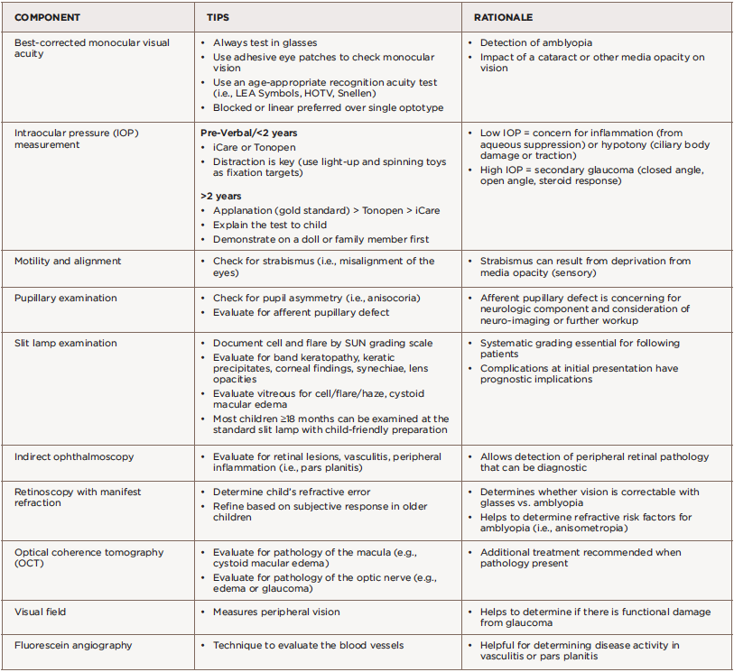
(click for larger image) Table 3: Eye Examination Components

Amblyopia is decreased vision in one or both eyes due to abnormal visual stimulation of the brain in infancy or early childhood. Uveitis can cause amblyopia via changes in refractive error from lenticular changes, glaucoma or deprivation secondary to media opacities such as band keratopathy, cataract, vitreous opacities and cystoid macular edema. Ancillary testing by visual field test (to measure peripheral vision), optical coherence tomography (to measure the thickness of the optic nerve and macula), and fundus photography may be performed based on clinical examination findings. This testing can help monitor complications of uveitis, such as glaucoma or cystoid macular edema.

ad goes here:advert-1
ADVERTISEMENT
SCROLL TO CONTINUE

Close collaboration between the rheumatologist and ophthalmologist is essential to optimize outcomes for the patient. The ophthalmologist closely monitors ocular inflammation through slit lamp exam and manages topical and peri/intraocular ophthalmic medications. The rheumatologist diagnoses and manages associated systemic diseases and treatment with systemic immunosuppressive medications.42 Regular communication is also important in establishing an overall treatment plan for each patient.

Unfortunately, when these evaluations are performed through specialists practicing in different healthcare systems, formal reports may not always be communicated between providers, creating fragmented care and potentially delayed diagnosis and treatment. Combined ophthalmology-rheumatology clinics can address these concerns. A review over a three-year period from a combined pediatric ophthal­mology and rheumatology clinic in Edinburgh, Scotland, suggested improved eye screening rates, improved convenience for both patients and providers, and prevention of delayed diagnosis and treatment in patients with uveitis present at their first eye exam.43

ad goes here:advert-2
ADVERTISEMENT
SCROLL TO CONTINUE

Our experience from the combined uveitis clinics at the Cincinnati Children’s Hospital Medical Center in Ohio has shown the advantages of subspecialty collaboration. Our clinic is staffed by both a pediatric ophthalmologist and a pediatric rheumatologist who perform evaluations simultaneously, which leads to a unified approach to care. The ophthalmologist performs an extensive eye assessment to determine the presence and extent of inflammation, and the rheumatologist conducts a full physical exam to evaluate for associated systemic conditions. The specialists can learn from one another, make treatment decisions based on their combined assessment and educate patient and families in real time if systemic therapies are required.

Page: 1 2 3 4 5 6 | Single Page
Share: 

Filed under:ConditionsRheumatoid Arthritis Tagged with:Juvenile Arthritis (JIA)Uveitis

Related Articles

    Watch Those Eyes

    December 1, 2007

    What you need to know about Uveitis in Rheumatic Diseases

    ARZTSAMUI / shutterstock.com

    Uveitis: A Brief Primer for the Rheumatologist

    November 12, 2020

    Uveitis is an umbrella term for intraocular inflammatory diseases that can lead to vision loss. It’s not just a concern for ophthalmologists. Uveitis occurs in approximately 2–5% of patients with inflammatory bowel disease, 6–9% of patients with psoriatic arthritis and 25% of patients with reactive arthritis. The prevalence may be as high as 33% in…

    New Guideline Recommends Frequent Monitoring & Collaboration for JIA-Associated Uveitis Management

    May 8, 2019

    As soon as pediatric patients are diagnosed with juvenile idiopathic arthritis (JIA), they should also be screened for uveitis, says ophthalmologist Gary Holland, MD. Otherwise, the University of California, Los Angeles, provider says, “Kids who are diagnosed with JIA may not come to an ophthalmologist until they have vision-limiting complications.” Uveitis is the most common…

    Oksana Kuzmina/shutterstock.comx

    Environmental Factors in Pediatric Systemic Autoimmune Diseases

    March 20, 2017

    Systemic autoimmune diseases are thought to result from immune dysregulation in genetically susceptible individuals who were exposed to environmental risk factors. Many studies have identified genetic risk factors for these diseases, but concordance rates among monozygotic twins are 25–40%, suggesting that nonheritable environmental factors play a more prominent role.1,2 Through carefully conducted epidemiologic and other…

  • About Us
  • Meet the Editors
  • Issue Archives
  • Contribute
  • Advertise
  • Contact Us
  • Copyright © 2025 by John Wiley & Sons, Inc. All rights reserved, including rights for text and data mining and training of artificial technologies or similar technologies. ISSN 1931-3268 (print). ISSN 1931-3209 (online).
  • DEI Statement
  • Privacy Policy
  • Terms of Use
  • Cookie Preferences