Video: Every Case Tells a Story| Webinar: ACR/CHEST ILD Guidelines in Practice

An official publication of the ACR and the ARP serving rheumatologists and rheumatology professionals

  • Conditions
    • Axial Spondyloarthritis
    • Gout and Crystalline Arthritis
    • Myositis
    • Osteoarthritis and Bone Disorders
    • Pain Syndromes
    • Pediatric Conditions
    • Psoriatic Arthritis
    • Rheumatoid Arthritis
    • Sjögren’s Disease
    • Systemic Lupus Erythematosus
    • Systemic Sclerosis
    • Vasculitis
    • Other Rheumatic Conditions
  • FocusRheum
    • ANCA-Associated Vasculitis
    • Axial Spondyloarthritis
    • Gout
    • Psoriatic Arthritis
    • Rheumatoid Arthritis
    • Systemic Lupus Erythematosus
  • Guidance
    • Clinical Criteria/Guidelines
    • Ethics
    • Legal Updates
    • Legislation & Advocacy
    • Meeting Reports
      • ACR Convergence
      • Other ACR meetings
      • EULAR/Other
    • Research Rheum
  • Drug Updates
    • Analgesics
    • Biologics/DMARDs
  • Practice Support
    • Billing/Coding
    • EMRs
    • Facility
    • Insurance
    • QA/QI
    • Technology
    • Workforce
  • Opinion
    • Patient Perspective
    • Profiles
    • Rheuminations
      • Video
    • Speak Out Rheum
  • Career
    • ACR ExamRheum
    • Awards
    • Career Development
  • ACR
    • ACR Home
    • ACR Convergence
    • ACR Guidelines
    • Journals
      • ACR Open Rheumatology
      • Arthritis & Rheumatology
      • Arthritis Care & Research
    • From the College
    • Events/CME
    • President’s Perspective
  • Search

Basics of Biologic Joint Reconstruction

Andreas Gomoll, MD  |  Issue: April 2012  |  April 6, 2012

There are no symptoms pathognomonic for cartilage defects, and patients are frequently diagnosed with meniscal tears or patellofemoral syndrome initially.

Physical Findings

ad goes here:advert-1
ADVERTISEMENT
SCROLL TO CONTINUE
Sagittal MRI
Figure 1: Sagittal MRI images of the medial femoral condyle of the same knee demonstrating substantial differences in sensitivity to detect cartilage defects with different sequences: (left) T1; (middle) proton density, fat saturated; (right) T2, fat saturated.

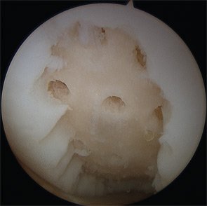
Similar to the patient’s presenting complaint, physical examination will not produce any findings specific for cartilage repair. Range of motion is usually preserved, although displaced osteochondral fragments can lead to intermittent locking. Large defects, especially in the patellofemoral compartment, can lead to reproducible catching or clicking with active and passive motion and patellar manipulation. Tenderness to palpation of the femoral condyles and joint spaces suggests the presence of synovitis. Given the importance of articular comorbidities, particular attention should be focused on the assessment of ligamentous stability, patellar maltracking, and alignment.

Diagnostic Imaging

Standard radiographs are utilized to screen for advanced osteoarthritic changes that would rule out cartilage repair as well as to assess the lower extremity for malalignment. Imaging includes weightbearing anteroposterior views in full extension; posterior–anterior (Rosenberg) views in mild flexion; lateral and patellofemoral views; and a full-length hip-to-ankle radiograph.

ad goes here:advert-2
ADVERTISEMENT
SCROLL TO CONTINUE

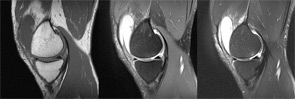
High-resolution (1.5 Tesla and greater) MRI is a reliable means to evaluate articular cartilage defects. The recent introduction of cartilage-specific imaging protocols has dramatically improved the quality of MRI scans and allows noninvasive monitoring after cartilage repair procedures (see Figure 1).16,17

Indications for Cartilage Repair

Osteochondral autograft transfer
Figure 2: Osteochondral autograft transfer for a defect of the lateral femoral condyle.

Microfracture of a large defect of the medial femoral condyle
Figure 3: Microfracture of a large defect of the medial femoral condyle.

Many cartilage defects are asymptomatic; therefore, careful assessment of other potential pain generators is critical prior to consideration of surgical treatment. Nonoperative treatment with rehabilitation, activity modification, and anti­inflammatory therapy should be considered for at least six months, especially for patellofemoral defects that typically respond well to a physical therapy regimen. Young patients with large defects are an exception to this recommendation, since lesion growth can occur even with symptomatic improvement from nonoperative treatment, potentially worsening the structural problem. For example, the majority of young patients experience almost complete resolution of knee pain following the removal of a loose osteochondritis dissecans (OCD) fragment; however, if the defect is left empty, 50% to 80% will develop radiographic evidence of osteoarthritis after eight to 10 years.18,19

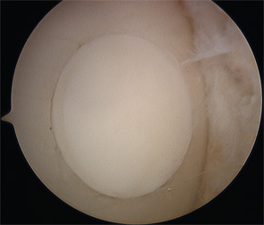
Surgical intervention is considered after establishing that the patient’s symptoms are consistent with a full-thickness (Outerbridge Grade 3 or 4) cartilage defect, and adequate nonoperative management has failed to provide acceptable pain relief. Extensive counseling about the long recovery period after cartilage repair is important to avoid unrealistic expectations and disappointment.

Page: 1 2 3 4 5 6 7 | Single Page
Share: 

Filed under:ConditionsOsteoarthritis and Bone Disorders Tagged with:bonecartilage repairimagingkneeOsteoarthritispatient careradiographrheumatologist

Related Articles
    HSCT for Severe Autoimmune Diseases

    HSCT for Severe Autoimmune Diseases

    August 14, 2017

    Despite the innovations of new biologics and disease-modifying anti-rheumatic drugs, a large unmet need remains for patients with rheumatic autoimmune disease. Treatment remains limited for many conditions, including for conditions with a dim prognosis, such as systemic sclerosis.1 One promising treatment avenue is hematopoietic stem-cell transplantation (HSCT). Here, we provide background on HSCT for severe…

    New Therapeutics for Osteoarthritis May Be in Sight

    April 1, 2015

    Overview of OA pathogenesis, recent discoveries suggest new treatment strategies are possible

    Does MRI Differentiate Osteoarthritis and Meniscal Tear in Knee Pain?

    December 19, 2017

    When a young patient arrives at a clinic complaining of knee pain with clicking or popping, a meniscal tear is often the culprit. “In young [people], there’s a pretty classic presentation of meniscal tear, with clicking and other mechanical symptoms, because the tear rubs up against different tissues,” says Jeffrey Katz, MD, MSc, a rheumatologist…

    Post-Traumatic Osteoarthritis: Managing OA That Develops After Joint Injuries & Reconstructive Surgery

    November 28, 2018

    CHICAGO—Joint trauma is one of many potential drivers of osteoarthritis disease activity and structural progression. In Post-Traumatic OA: Pathogenesis, Clinical Evolution and Management, a session at the 2018 ACR/ARHP Annual Meeting, experts discussed the effects of sports and other injuries on even young patients’ joints. Post-traumatic osteoarthritis (OA) may account for 12% of hip, knee…

  • About Us
  • Meet the Editors
  • Issue Archives
  • Contribute
  • Advertise
  • Contact Us
  • Copyright © 2025 by John Wiley & Sons, Inc. All rights reserved, including rights for text and data mining and training of artificial technologies or similar technologies. ISSN 1931-3268 (print). ISSN 1931-3209 (online).
  • DEI Statement
  • Privacy Policy
  • Terms of Use
  • Cookie Preferences