Video: Every Case Tells a Story| Webinar: ACR/CHEST ILD Guidelines in Practice

An official publication of the ACR and the ARP serving rheumatologists and rheumatology professionals

  • Conditions
    • Axial Spondyloarthritis
    • Gout and Crystalline Arthritis
    • Myositis
    • Osteoarthritis and Bone Disorders
    • Pain Syndromes
    • Pediatric Conditions
    • Psoriatic Arthritis
    • Rheumatoid Arthritis
    • Sjögren’s Disease
    • Systemic Lupus Erythematosus
    • Systemic Sclerosis
    • Vasculitis
    • Other Rheumatic Conditions
  • FocusRheum
    • ANCA-Associated Vasculitis
    • Axial Spondyloarthritis
    • Gout
    • Psoriatic Arthritis
    • Rheumatoid Arthritis
    • Systemic Lupus Erythematosus
  • Guidance
    • Clinical Criteria/Guidelines
    • Ethics
    • Legal Updates
    • Legislation & Advocacy
    • Meeting Reports
      • ACR Convergence
      • Other ACR meetings
      • EULAR/Other
    • Research Rheum
  • Drug Updates
    • Analgesics
    • Biologics/DMARDs
  • Practice Support
    • Billing/Coding
    • EMRs
    • Facility
    • Insurance
    • QA/QI
    • Technology
    • Workforce
  • Opinion
    • Patient Perspective
    • Profiles
    • Rheuminations
      • Video
    • Speak Out Rheum
  • Career
    • ACR ExamRheum
    • Awards
    • Career Development
  • ACR
    • ACR Home
    • ACR Convergence
    • ACR Guidelines
    • Journals
      • ACR Open Rheumatology
      • Arthritis & Rheumatology
      • Arthritis Care & Research
    • From the College
    • Events/CME
    • President’s Perspective
  • Search

Basics of Biologic Joint Reconstruction

Andreas Gomoll, MD  |  Issue: April 2012  |  April 6, 2012

Patients who smoke, are obese (BMI >35), or carry a diagnosis of inflammatory arthritis are not good candidates for cartilage repair surgery.20 Advanced degenerative changes with more than 50% joint space narrowing are considered a contraindication to cartilage repair in all but the very young who have intolerable symptoms and no other reasonable therapeutic options.

Treatment Algorithm

Once a decision has been made to proceed with cartilage repair, a surgical plan is formulated that defines not only the specific repair technique best suited to address the specific cartilage defect, but also incorporates any procedures required to remedy potential articular comorbidities. Treatment strategies for cartilage repair are based primarily on defect location and size. The two most common locations for cartilage defects are on the (medial) femoral condyle and in the patellofemoral compartment, each with their own treatment algorithms.1,21,22

ad goes here:advert-1
ADVERTISEMENT
SCROLL TO CONTINUE

Tibiofemoral Compartment

The choice of cartilage repair procedure for femoral defects is primarily dictated by defect size. Generally, the less invasive procedures of microfracture and osteochondral autograft transfer (OAT) have demonstrated acceptable outcomes only in defects smaller than approximately 3–4 cm2. Larger defects demonstrated only transient improvement in function, or increased donor site morbidity, respectively.23-26 Both microfracture and OAT consistently produce good and excellent results in 60% to 80% of patients with small lesions (<2–4 cm2) on the femoral condyles.27-30

Larger defects therefore are mostly treated with more invasive procedures such as osteochondral allograft transplantation or autologous chondrocyte implantation (ACI). While no studies have been performed to directly compare these more invasive procedures, large cohort studies have demonstrated good and excellent results in more than 70% of patients for each.3,31-35 Generally, defects limited to the articular surface are best treated with ACI, while defects with associated abnormalities of the subchondral bone should be considered for osteochondral allograft transplantation, which replaces both cartilage and the underlying bone.

ad goes here:advert-2
ADVERTISEMENT
SCROLL TO CONTINUE

Patellofemoral Compartment

Cartilage defect
Figure 4: Cartilage defect with delamination on the medial femoral condyle (left). After placement of a fresh osteochondral allograft (right).

The patellofemoral (PF) compartment is a difficult location for cartilage repair, and all techniques do worse here compared with the tibiofemoral compartment, due to its more complex geometry and higher shear forces. The importance of correcting adverse factors, such as patellar maltracking, is particularly important in this location.

While microfracture, OAT, and osteochondral allograft have generally good outcomes in the femoral condyles, there is a growing consensus that they should be used cautiously in the patellofemoral compartment except in very specific cases.

Surgical Procedures to Treat Cartilage Defects

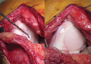
Debridement and chondroplasty (removal of degenerated tissue and creation of mechanically stable cartilage with vertical shoulders) is generally performed arthroscopically. This can be the definitive treatment for incidentally found lesions, or initial treatment for symptomatic defects prior to consideration of more invasive cartilage repair procedures. Patients recover quickly after a debridement and chondroplasty, and patients can be fully weight bearing, but should allow pain and swelling to resolve prior to returning to activities.

Page: 1 2 3 4 5 6 7 | Single Page
Share: 

Filed under:ConditionsOsteoarthritis and Bone Disorders Tagged with:bonecartilage repairimagingkneeOsteoarthritispatient careradiographrheumatologist

Related Articles
    HSCT for Severe Autoimmune Diseases

    HSCT for Severe Autoimmune Diseases

    August 14, 2017

    Despite the innovations of new biologics and disease-modifying anti-rheumatic drugs, a large unmet need remains for patients with rheumatic autoimmune disease. Treatment remains limited for many conditions, including for conditions with a dim prognosis, such as systemic sclerosis.1 One promising treatment avenue is hematopoietic stem-cell transplantation (HSCT). Here, we provide background on HSCT for severe…

    New Therapeutics for Osteoarthritis May Be in Sight

    April 1, 2015

    Overview of OA pathogenesis, recent discoveries suggest new treatment strategies are possible

    Does MRI Differentiate Osteoarthritis and Meniscal Tear in Knee Pain?

    December 19, 2017

    When a young patient arrives at a clinic complaining of knee pain with clicking or popping, a meniscal tear is often the culprit. “In young [people], there’s a pretty classic presentation of meniscal tear, with clicking and other mechanical symptoms, because the tear rubs up against different tissues,” says Jeffrey Katz, MD, MSc, a rheumatologist…

    Post-Traumatic Osteoarthritis: Managing OA That Develops After Joint Injuries & Reconstructive Surgery

    November 28, 2018

    CHICAGO—Joint trauma is one of many potential drivers of osteoarthritis disease activity and structural progression. In Post-Traumatic OA: Pathogenesis, Clinical Evolution and Management, a session at the 2018 ACR/ARHP Annual Meeting, experts discussed the effects of sports and other injuries on even young patients’ joints. Post-traumatic osteoarthritis (OA) may account for 12% of hip, knee…

  • About Us
  • Meet the Editors
  • Issue Archives
  • Contribute
  • Advertise
  • Contact Us
  • Copyright © 2025 by John Wiley & Sons, Inc. All rights reserved, including rights for text and data mining and training of artificial technologies or similar technologies. ISSN 1931-3268 (print). ISSN 1931-3209 (online).
  • DEI Statement
  • Privacy Policy
  • Terms of Use
  • Cookie Preferences