Video: Every Case Tells a Story| Webinar: ACR/CHEST ILD Guidelines in Practice

An official publication of the ACR and the ARP serving rheumatologists and rheumatology professionals

  • Conditions
    • Axial Spondyloarthritis
    • Gout and Crystalline Arthritis
    • Myositis
    • Osteoarthritis and Bone Disorders
    • Pain Syndromes
    • Pediatric Conditions
    • Psoriatic Arthritis
    • Rheumatoid Arthritis
    • Sjögren’s Disease
    • Systemic Lupus Erythematosus
    • Systemic Sclerosis
    • Vasculitis
    • Other Rheumatic Conditions
  • FocusRheum
    • ANCA-Associated Vasculitis
    • Axial Spondyloarthritis
    • Gout
    • Psoriatic Arthritis
    • Rheumatoid Arthritis
    • Systemic Lupus Erythematosus
  • Guidance
    • Clinical Criteria/Guidelines
    • Ethics
    • Legal Updates
    • Legislation & Advocacy
    • Meeting Reports
      • ACR Convergence
      • Other ACR meetings
      • EULAR/Other
    • Research Rheum
  • Drug Updates
    • Analgesics
    • Biologics/DMARDs
  • Practice Support
    • Billing/Coding
    • EMRs
    • Facility
    • Insurance
    • QA/QI
    • Technology
    • Workforce
  • Opinion
    • Patient Perspective
    • Profiles
    • Rheuminations
      • Video
    • Speak Out Rheum
  • Career
    • ACR ExamRheum
    • Awards
    • Career Development
  • ACR
    • ACR Home
    • ACR Convergence
    • ACR Guidelines
    • Journals
      • ACR Open Rheumatology
      • Arthritis & Rheumatology
      • Arthritis Care & Research
    • From the College
    • Events/CME
    • President’s Perspective
  • Search

Case Report: A Behçet’s Patient Develops Cerebral Venous Sinus Thrombi

Javaneh Lyons, MD, MSc, Alana Nevares, MD  |  Issue: December 2018  |  December 17, 2018

A 39-year-old woman presented at the emergency department with three weeks of progressive, constant and pulsatile right-sided headache. She said her headache was worse in the morning and when she would bend forward. She reported associated nausea and vomiting.

On initial assessment, she did not have any focal neurological deficits. Her medical history was significant for tobacco use, alcohol abuse, depression and recent diagnosis of Behçet’s disease. Behçet’s was diagnosed on the basis of many years of recurrent oral ulcers, one year of recurrent genital ulcers, diffuse arthralgia, positive pathergy, history suggestive of prior iritis and pustular skin eruption with an otherwise negative rheumatologic workup. At the time of presentation, she was not on any treatment for Behçet’s disease due to financial limitations.

ad goes here:advert-1
ADVERTISEMENT
SCROLL TO CONTINUE

In the emergency department, she was promptly evaluated with magnetic resonance imaging (MRI) and magnetic resonance venography (MRV), and was found to have extensive sinus thrombosis involving the right internal jugular vein, right sigmoid sinus, right transverse sinus and right superior sagittal sinus (see Figures 1 and 2). She did not have any findings suggestive of infarction or hemorrhage.

Her ophthalmologic evaluation did not show any evidence of subclinical uveitis or optic neuritis. The findings of cerebral venous sinus thrombi (CVST) were attributed to Behçet’s disease, and a hypercoagulable workup, including factor V Leiden, prothrombin 20210 mutation and antiphospholipid antibodies, was later unrevealing.

ad goes here:advert-2
ADVERTISEMENT
SCROLL TO CONTINUE

Treatment with anticoagulation and immunosuppression was initiated. She received high-dose prednisone (1 mg/kg) and was started on azathioprine (1 mg/kg; 50 mg twice daily), with resolution of her headache within the first two weeks of treatment. Prednisone was gradually tapered over 12 months without signs or symptoms concerning for relapse. For anticoagulation, she initially received unfractionated heparin and was then transitioned to warfarin to complete a total of eight months of treatment. The duration of anticoagulation was determined on the basis of resolution of the thrombi on subsequent imaging at three and six months.

Figure 1: Axial T1 contrast MR image showing non-enhancement within the (A) right transverse sinus, (B) right sigmoid sinus and (C) right internal jugular vein. Associated dural enhancement is visualized around the transverse sinus (A) and sigmoid sinus (B).

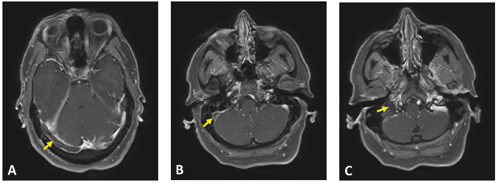
(click for larger image) Figure 1: Axial T1 contrast MR image showing non-enhancement within the (A) right transverse sinus, (B) right sigmoid sinus and (C) right internal jugular vein. Associated dural enhancement is visualized around the transverse sinus (A) and sigmoid sinus (B).

Behçet Disease

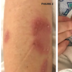
Behçet’s disease was first described by Hulusi Behçet, a Turkish dermatologist, in 1937 and was defined as a syndrome of recurrent oral aphthous ulcers, genital ulcers and uveitis.1 It’s classified as a variable vessel vasculitis (can affect vessels of any size and type), affecting both arteries and veins.2 The definition of Behçet’s disease has been refined over the years and the most widely used diagnostic criteria today are based on the International Study Group Diagnostic Criteria published in 1990. These include recurrent oral ulcerations of at least three incidents in a 12-month period, in addition to two other manifestations of recurrent genital ulcers, eye lesions, skin lesions or a positive pathergy test.1,3

In 2014, classification criteria were revised by the International Criteria for Behçet’s Disease (ICBD), which increased their sensitivity by including neurological and vascular manifestations in addition to oral and genital ulcerations. A score of 4 or more points is estimated to have 93.9% sensitivity and 92.1% specificity.4

Page: 1 2 3 4 5 | Single Page
Share: 

Filed under:ConditionsVasculitis Tagged with:azathioprineBehçet’s diseasecase reportcerebral venous sinus thrombi (CVST)

Related Articles

    Case Report: An Unusual Presentation of Neuro-Behçet’s Disease

    April 15, 2022

    A 44-year-old woman presented to the emergency department with bifrontal headaches that had started approximately one month earlier. She was diagnosed with migraines and discharged home. Three days later, the patient returned to the emergency department upon recurrence of her headaches, and this time she also reported abnormal leg movements. A computerized tomography (CT) scan…

    Submassive pulmonary embolism. Filling defects seen in the pulmonary artery.

    Case Report Illustrates Hypercoagulability in Behçet’s Disease

    June 21, 2018

    Behçet’s disease is a chronic, relapsing and remitting vasculitis with multisystem involvement. Commonly referred to as the Silk Road disease due to its prevalence in the Asian and Mediterranean region of the traditional Silk Road, Behçet’s was first described by Hippocrates as a triad of symptoms—genital and oral ulcers with uveitis—and attributed to links with…

    Case Report: Which Vasculitis Is It?

    June 15, 2020

    A 13-year-old, adopted girl of unknown ancestry with social anxiety, selective mutism and Takayasu arteritis presented for evaluation of severe, painful, gingival hyperplasia, which limited her oral intake and resulted in weight loss. The young patient was diagnosed with Takayasu arteritis at age 8, when she presented with a persistently elevated erythrocyte sedimentation rate (ESR),…

    Tracking Patient Manifestations of Behçet’s Syndrome around the World

    July 1, 2010

    Tracking patient manifestations of Behçet’s syndrome around the world

  • About Us
  • Meet the Editors
  • Issue Archives
  • Contribute
  • Advertise
  • Contact Us
  • Copyright © 2025 by John Wiley & Sons, Inc. All rights reserved, including rights for text and data mining and training of artificial technologies or similar technologies. ISSN 1931-3268 (print). ISSN 1931-3209 (online).
  • DEI Statement
  • Privacy Policy
  • Terms of Use
  • Cookie Preferences