Video: Every Case Tells a Story| Webinar: ACR/CHEST ILD Guidelines in Practice

An official publication of the ACR and the ARP serving rheumatologists and rheumatology professionals

  • Conditions
    • Axial Spondyloarthritis
    • Gout and Crystalline Arthritis
    • Myositis
    • Osteoarthritis and Bone Disorders
    • Pain Syndromes
    • Pediatric Conditions
    • Psoriatic Arthritis
    • Rheumatoid Arthritis
    • Sjögren’s Disease
    • Systemic Lupus Erythematosus
    • Systemic Sclerosis
    • Vasculitis
    • Other Rheumatic Conditions
  • FocusRheum
    • ANCA-Associated Vasculitis
    • Axial Spondyloarthritis
    • Gout
    • Psoriatic Arthritis
    • Rheumatoid Arthritis
    • Systemic Lupus Erythematosus
  • Guidance
    • Clinical Criteria/Guidelines
    • Ethics
    • Legal Updates
    • Legislation & Advocacy
    • Meeting Reports
      • ACR Convergence
      • Other ACR meetings
      • EULAR/Other
    • Research Rheum
  • Drug Updates
    • Analgesics
    • Biologics/DMARDs
  • Practice Support
    • Billing/Coding
    • EMRs
    • Facility
    • Insurance
    • QA/QI
    • Technology
    • Workforce
  • Opinion
    • Patient Perspective
    • Profiles
    • Rheuminations
      • Video
    • Speak Out Rheum
  • Career
    • ACR ExamRheum
    • Awards
    • Career Development
  • ACR
    • ACR Home
    • ACR Convergence
    • ACR Guidelines
    • Journals
      • ACR Open Rheumatology
      • Arthritis & Rheumatology
      • Arthritis Care & Research
    • From the College
    • Events/CME
    • President’s Perspective
  • Search

Case Report: A Non-Tuberculous Mycobacterial Infection

Bradley Bohman, MD, & Jawad Bilal, MBBS  |  Issue: January 2021  |  January 20, 2021

Magnetic resonance imaging of the patient’s left knee shows joint effusion, synovial thickening, and periarticular muscle and soft tissue edema.

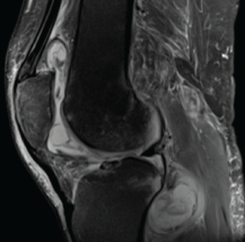
Figure 1. Magnetic resonance imaging of the patient’s left knee shows joint effusion, synovial thickening, and periarticular muscle and soft tissue edema.

Tumor necrosis factor-α inhibitors (TNFi’s) have emerged as an integral part of therapeutic strategies for several rheumatic diseases. TNF-α is a pro-inflammatory cytokine implicated in the pathogenesis of rheumatoid arthritis (RA), seronegative spondyloarthropathies and inflammatory bowel disease (IBD). It also plays a central role in the immune response to mycobacterial infection. 

Many biologic agents, particularly TNFi’s, are associated with an increased risk of tuberculosis; however, the risk of non-tuberculous mycobacterial infections among patients treated with a TNFi has not been studied extensively. 

ad goes here:advert-1
ADVERTISEMENT
SCROLL TO CONTINUE

We present this report on a patient with sarcoidosis receiving TNFi therapy who developed a disseminated non-tuberculous mycobacterial infection.

Case History

A 69-year-old man presented to the clinic with symptoms of progressively worsening left knee pain, stiffness and swelling over six months. He was unable to bear weight on the affected leg and was wheelchair bound. He had previously been diagnosed with both neurosarcoidosis and osteo­arthritis. He had initially been treated with glucocorticoid monotherapy. Because he deteriorated clinically, 400 mg of intra­venous (IV) infliximab every six weeks and 500 mg of mycophenolate mofetil twice a day orally were added to his medical regimen.

ad goes here:advert-2
ADVERTISEMENT
SCROLL TO CONTINUE

His knee had initially been evaluated by his primary care provider; plain radiographs of the knees demonstrated osteo­arthritis. Subsequently, he was referred to a sports medicine specialist and an orthopedic surgeon for further evaluation. Oral analgesics, intra-articular steroid injection and physical therapy provided him with no relief, and he was referred to our division for further evaluation.

In our clinic, he was afebrile; his blood pressure was 152/82 mmHg, and his heart rate was 66 bpm. His left knee was swollen, and both passive and active movements were limited by pain.

Laboratory tests on his initial presentation demonstrated leukocytosis; his white blood cell count was 13,200/uL with 86% neutrophils. His complete metabolic panel and urinalysis were unremarkable. Rheum­atoid factor, anti-cyclic citrullinated protein antibodies and anti-double stranded DNA antibodies were not detected on immunological studies. Tests for Borrelia and Coccidioides IgG and IgM antibodies were negative. 

Magnetic resonance imaging (MRI) of the left knee joint revealed a moderate joint effusion with diffuse synovial thickening, and periarticular muscle and soft tissue edema (see Figure 1, above). 

Synovial fluid aspirate demonstrated 8,935 nucleated cells (70% mononuclear) with Mycobacterium avium-intra­cellulare growth in modified Middlebrook 7H9 broth, a liquid growth medium used to culture Mycobacterium. 

Infliximab was stopped, and the patient was treated with ethambutol and azithromycin.

Over the next month, the patient noticed low-grade fever, malaise, night sweats and unintentional weight loss of 10 lbs. He underwent open synovectomy of the left knee. The synovial tissue demonstrated an active granulomatous synovitis (see Figure 2, and synovial cultures grew Mycobacterium avium complex (MAC). 

Page: 1 2 3 4 | Single Page
Share: 

Filed under:ConditionsOther Rheumatic Conditions Tagged with:bacteriacase reportInfectiontumor necrosis factor inhibitor (TNFi)

Related Articles

    How Tuberculosis Has Shaped Medicine and Society

    May 17, 2017

    Pathologists are legendary for blending their work product with the culinary arts. Through the years, their use of delectable foods as descriptors has created a clever way to indelibly link in the minds of clinicians the histopathologic observations of disease with an assortment of these tasty foods: There is the depiction of an apple green…

    Case Report: A Mycobacterium kansasii Infection

    May 13, 2021

    A 61-year-old white woman presented to our rheumatology clinic in New England to establish care in early June 2018, following a move from Texas. She reported a medical history of inflammatory bowel disease, uveitis and sero­negative inflammatory arthritis, which was difficult to control and required the use of multiple medications. At her initial visit, she…

    Rheumatology Drugs at a Glance, Part 3: Rheumatoid Arthritis

    August 16, 2019

    Over the past few years, bio­similars and other new drugs have been introduced to treat rheumatic illnesses. Some of the conditions we treat have numerous drug options, others have few or only off-label options. This series, “Rheumatology Drugs at a Glance,” provides streamlined information on the administration of biologic, biosimilar and small molecule inhibitor drugs…

    Drug Reduction Strategies, Disease Control for Patients with RA in Remission

    April 20, 2017

    WASHINGTON, D.C.—Clinical aspects of managing patients with rheumatoid arthritis (RA) in remission were discussed by a panel of experts at the 2016 ACR/ARHP Annual Meeting during the session titled Rheumatoid Arthritis—Clinical Aspects IV: Managing Patients in Remission. Among the issues raised were strategies to taper or discontinue biologic therapies, as well as clinical predictors of…

  • About Us
  • Meet the Editors
  • Issue Archives
  • Contribute
  • Advertise
  • Contact Us
  • Copyright © 2025 by John Wiley & Sons, Inc. All rights reserved, including rights for text and data mining and training of artificial technologies or similar technologies. ISSN 1931-3268 (print). ISSN 1931-3209 (online).
  • DEI Statement
  • Privacy Policy
  • Terms of Use
  • Cookie Preferences