Video: Every Case Tells a Story| Webinar: ACR/CHEST ILD Guidelines in Practice

An official publication of the ACR and the ARP serving rheumatologists and rheumatology professionals

  • Conditions
    • Axial Spondyloarthritis
    • Gout and Crystalline Arthritis
    • Myositis
    • Osteoarthritis and Bone Disorders
    • Pain Syndromes
    • Pediatric Conditions
    • Psoriatic Arthritis
    • Rheumatoid Arthritis
    • Sjögren’s Disease
    • Systemic Lupus Erythematosus
    • Systemic Sclerosis
    • Vasculitis
    • Other Rheumatic Conditions
  • FocusRheum
    • ANCA-Associated Vasculitis
    • Axial Spondyloarthritis
    • Gout
    • Psoriatic Arthritis
    • Rheumatoid Arthritis
    • Systemic Lupus Erythematosus
  • Guidance
    • Clinical Criteria/Guidelines
    • Ethics
    • Legal Updates
    • Legislation & Advocacy
    • Meeting Reports
      • ACR Convergence
      • Other ACR meetings
      • EULAR/Other
    • Research Rheum
  • Drug Updates
    • Analgesics
    • Biologics/DMARDs
  • Practice Support
    • Billing/Coding
    • EMRs
    • Facility
    • Insurance
    • QA/QI
    • Technology
    • Workforce
  • Opinion
    • Patient Perspective
    • Profiles
    • Rheuminations
      • Video
    • Speak Out Rheum
  • Career
    • ACR ExamRheum
    • Awards
    • Career Development
  • ACR
    • ACR Home
    • ACR Convergence
    • ACR Guidelines
    • Journals
      • ACR Open Rheumatology
      • Arthritis & Rheumatology
      • Arthritis Care & Research
    • From the College
    • Events/CME
    • President’s Perspective
  • Search

Case Report: A Non-Tuberculous Mycobacterial Infection

Bradley Bohman, MD, & Jawad Bilal, MBBS  |  Issue: January 2021  |  January 20, 2021

Synovium with granulomatous inflammation consisting of aggregates of histiocytes and multinucleated giant cells, with surrounding lymphocytes and plasma cells. Sections contain no identifiable organisms or crystal deposition. (GMS and AFB immunohistochemical stains were negative.) H&E, 100x. Source: Picture and description by Mary Hansen Smith MD, University of Arizona, Department of Pathology.

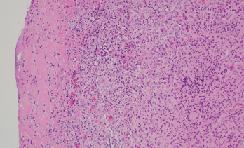
Figure 2. Synovium with granulomatous inflammation consisting of aggregates of histiocytes and multinucleated giant cells, with surrounding lymphocytes and plasma cells. Sections contain no identifiable organisms or crystal deposition. (GMS and AFB immunohistochemical stains were negative.) H&E, 100x.
Source: Picture and description by Mary Hansen Smith MD, University of Arizona, Department of Pathology.

Rifampin was added to his antibiotic regimen. Further evaluation included a high-resolution computed tomography (HRCT) of the chest, which demonstrated sub-centimeter scattered nodules (see Figure 3). Given the nodularity, a diagnosis of disseminated MAC
was established. 

The patient’s neurosarcoidosis initially remained in remission, but two years later, he developed a cervical cord lesion. The patient was started back on infliximab monotherapy while continuing to receive treatment with azithromycin, ethambutol and rifampin. 

ad goes here:advert-1
ADVERTISEMENT
SCROLL TO CONTINUE

After three doses of infliximab, the patient developed increasing spasticity in the bilateral lower extremities, decreased ability to move his legs and weakness of the right arm. The patient was transitioned from infliximab to rituximab.1 The patient received four weekly doses of 375 mg/m2 of rituximab without discernable benefit. Later, he received radiation to the cervical spine for refractory neurosarcoidosis, but continued to experience a decline in strength. 

The patient then started treatment with 300 mg of IV ocrelizumab and 250 mg of IV methylprednisolone. He was continued on triple therapy (i.e., azithromycin, ethambutol and rifampin) to prevent recurrence. The patient continues to follow up with his pulmonologist and neurologist, who are monitoring his response to therapy.

ad goes here:advert-2
ADVERTISEMENT
SCROLL TO CONTINUE

Discussion

Individuals being treated with immuno­suppressive agents are more susceptible to fungal and mycobacterial infections. The currently available evidence suggests an increased risk of Mycobacterium tuber­culosis infection with the use of TNFi’s.2,3 However, the occurrence of non-tuberculous mycobacterial infections with the use of biologics is not well established.

In a case-control study, of 30,772 patients receiving a TNFi, 158 (0.51%) developed targeted fungal and/or mycobacterial infections. Non-tuberculous mycobacterial infections were identified in 11% of these patients. The study also looked at the odds ratio for patients taking different TNFi’s compared with etanercept to evaluate how likely a patient would be treated for a fungal or mycobacterial infection. For adalimumab vs. etanercept, the odds ratio was 1.05 (0.69–1.62), infliximab vs. etanercept 1.08 (0.66–1.77), and for certolizumab vs. etanercept 0.78 (0.07–8.21). Thus, the study did not see a statistically significant difference between different TNFi’s when compared to etanercept.4 

High-resolution computed tomography of the chest demonstrated sub-centimeter, scattered nodularity consistent with disseminated Mycobacterium avium complex.

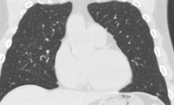
Figure 3. High-resolution computed tomography of the chest demonstrated sub-centimeter, scattered nodularity consistent with disseminated Mycobacterium avium complex.

A retrospective cohort study involving 42,180 rheumatoid arthritis patients suggested the occurrence of Mycobacterium tuberculosis varied with different biologic therapies. The hazard ratio for adalimumab was 1.52 (1.18–1.96), etanercept 1.16 (0.9–1.41) and rituximab 0.08 (0.02–0.31) using conventional synthetic disease-modifying anti-rheumatic drugs-exposed patients as a reference group.5 

Another retrospective cohort study involving 951 patients evaluated the occurrence of tuberculosis in RA patients on biologics. The findings suggested a higher Mycobacterium tuberculosis risk for patients receiving etanercept and adalimumab, but not for those receiving golimumab, abatacept, tocilizumab or tofacitinib.6

A retrospective study was conducted to identify the occurrence of non-tuberculous mycobacterial and tuberculosis cases among 8,418 patients with inflammatory diseases who received anti-TNF therapy. The occurrence of Mycobacterium tuberculosis and non-tuberculous mycobacterial disease was found to be fivefold higher in those receiving a TNFi than in those not receiving a TNFi and tenfold more elevated than in the general population. Non-tuberculous mycobacterial cases occurred more frequently among patients taking a TNFi, and interestingly, these patients were more likely to have chronic lung disease and gastroesophageal reflux disease.7

Page: 1 2 3 4 | Single Page
Share: 

Filed under:ConditionsOther Rheumatic Conditions Tagged with:bacteriacase reportInfectiontumor necrosis factor inhibitor (TNFi)

Related Articles

    How Tuberculosis Has Shaped Medicine and Society

    May 17, 2017

    Pathologists are legendary for blending their work product with the culinary arts. Through the years, their use of delectable foods as descriptors has created a clever way to indelibly link in the minds of clinicians the histopathologic observations of disease with an assortment of these tasty foods: There is the depiction of an apple green…

    Case Report: A Mycobacterium kansasii Infection

    May 13, 2021

    A 61-year-old white woman presented to our rheumatology clinic in New England to establish care in early June 2018, following a move from Texas. She reported a medical history of inflammatory bowel disease, uveitis and sero­negative inflammatory arthritis, which was difficult to control and required the use of multiple medications. At her initial visit, she…

    Rheumatology Drugs at a Glance, Part 3: Rheumatoid Arthritis

    August 16, 2019

    Over the past few years, bio­similars and other new drugs have been introduced to treat rheumatic illnesses. Some of the conditions we treat have numerous drug options, others have few or only off-label options. This series, “Rheumatology Drugs at a Glance,” provides streamlined information on the administration of biologic, biosimilar and small molecule inhibitor drugs…

    Drug Reduction Strategies, Disease Control for Patients with RA in Remission

    April 20, 2017

    WASHINGTON, D.C.—Clinical aspects of managing patients with rheumatoid arthritis (RA) in remission were discussed by a panel of experts at the 2016 ACR/ARHP Annual Meeting during the session titled Rheumatoid Arthritis—Clinical Aspects IV: Managing Patients in Remission. Among the issues raised were strategies to taper or discontinue biologic therapies, as well as clinical predictors of…

  • About Us
  • Meet the Editors
  • Issue Archives
  • Contribute
  • Advertise
  • Contact Us
  • Copyright © 2025 by John Wiley & Sons, Inc. All rights reserved, including rights for text and data mining and training of artificial technologies or similar technologies. ISSN 1931-3268 (print). ISSN 1931-3209 (online).
  • DEI Statement
  • Privacy Policy
  • Terms of Use
  • Cookie Preferences