Video: Every Case Tells a Story| Webinar: ACR/CHEST ILD Guidelines in Practice

An official publication of the ACR and the ARP serving rheumatologists and rheumatology professionals

  • Conditions
    • Axial Spondyloarthritis
    • Gout and Crystalline Arthritis
    • Myositis
    • Osteoarthritis and Bone Disorders
    • Pain Syndromes
    • Pediatric Conditions
    • Psoriatic Arthritis
    • Rheumatoid Arthritis
    • Sjögren’s Disease
    • Systemic Lupus Erythematosus
    • Systemic Sclerosis
    • Vasculitis
    • Other Rheumatic Conditions
  • FocusRheum
    • ANCA-Associated Vasculitis
    • Axial Spondyloarthritis
    • Gout
    • Psoriatic Arthritis
    • Rheumatoid Arthritis
    • Systemic Lupus Erythematosus
  • Guidance
    • Clinical Criteria/Guidelines
    • Ethics
    • Legal Updates
    • Legislation & Advocacy
    • Meeting Reports
      • ACR Convergence
      • Other ACR meetings
      • EULAR/Other
    • Research Rheum
  • Drug Updates
    • Analgesics
    • Biologics/DMARDs
  • Practice Support
    • Billing/Coding
    • EMRs
    • Facility
    • Insurance
    • QA/QI
    • Technology
    • Workforce
  • Opinion
    • Patient Perspective
    • Profiles
    • Rheuminations
      • Video
    • Speak Out Rheum
  • Career
    • ACR ExamRheum
    • Awards
    • Career Development
  • ACR
    • ACR Home
    • ACR Convergence
    • ACR Guidelines
    • Journals
      • ACR Open Rheumatology
      • Arthritis & Rheumatology
      • Arthritis Care & Research
    • From the College
    • Events/CME
    • President’s Perspective
  • Search

Case Report: A Rare Mimic of Giant Cell Arteritis

Iryna Nemesh, MD, Saleema Kherani, MD MPH, Shikha Singla, MD, & William Wirostko, MD  |  Issue: November 2021  |  November 14, 2021

Spectral domain optical coherence tomography (SD-OCT) of the left eye with hyper-reflective retinal pigment epithelial (RPE) nodularity (white arrow) and overlying loss of normal photo receptor architecture.

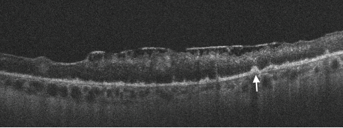
Figure 2. Spectral domain optical coherence tomography (SD-OCT) of the left eye with hyper-reflective retinal pigment epithelial (RPE) nodularity (white arrow) and overlying loss of normal photo receptor architecture.

Discussion

Ocular syphilis is rare and tends to occur at any stage of the disease. However, it is commonly associated with neurosyphilis. Its incidence has been increasing in the developed countries for the past few years. This may be related in part to decreased condom use in high-risk populations following the advent of highly active anti-retroviral therapy (HAART).5

Because its clinical manifestations vary, ocular syphilis is a big masquerader and may be challenging to diagnose. Ocular symptoms are commonly non-specific and may lead to a delay in diagnosis and treatment. New-onset, monocular, blurry vision is usually an early manifestation; however, other non-specific systemic symptoms can mask the diagnosis.

ad goes here:advert-1
ADVERTISEMENT
SCROLL TO CONTINUE

The clinical presentation of monocular vision changes when present with relevant associated symptoms can be concerning for GCA, which can lead to permanent visual loss if untreated. Therefore establishing a definitive diagnosis is paramount.

Elevated inflammatory markers may be present in both conditions. Patients with such a presentation are often empirically treated with high-dose systemic steroids and can be subjected to a temporal artery biopsy. Because the diagnostic yield of a temporal artery biopsy is low, these patients can remain on treatment despite a negative biopsy result. A diagnosis of ocular syphilis in such cases can be missed, resulting in permanent visual impairment. A timely diagnosis of this important clinical entity can mitigate unnecessary testing and prevent complications.

ad goes here:advert-2
ADVERTISEMENT
SCROLL TO CONTINUE

Based on our experience, we recommend testing for syphilis and tuberculosis prior to initiating systemic steroid therapy. Additionally, a detailed sexual history must be obtained, and those with high-risk behaviors must also be tested for syphilis.


Iryna Nemesh, MD, is a rheumatology fellow in the Department of Medicine, Division of Rheumatology, Medical College of Wisconsin Affiliated Hospitals, Milwaukee.

Saleema Kherani, MD, MPH, is an ophthalmology resident in the Department of Ophthalmology, Medical College of Wisconsin Affiliated Hospitals, Milwaukee.

Shikha Singla, MD, is an assistant professor in the Department of Medicine, Division of Rheumatology, Medical College of Wisconsin Affiliated Hospitals, Milwaukee.

William Wirostko, MD, is a professor of vitreoretinal surgery and ocular oncology in the Department of Ophthalmology, Medical College of Wisconsin Affiliated Hospitals, Milwaukee.

References

  1. Doris JP, Saha K, Jones NP, Sukthankar A. Ocular syphilis: The new epidemic. Eye (Lond). 2006 Jun;20(6):703–705.
  2. Majumder PD, Chen EJ, Shah J, et al. Ocular syphilis: An update. Ocul Immunol Inflamm. 2019;27(1):117–125.
  3. Kiss S, Damico FM, Young LH. Ocular manifestations and treatment of syphilis. Semin Ophthalmol. 2005;20(3):161–167.
  4. Biousse V, Newman NJ. Ischemic optic neuropathies. N Engl J Med. 2015 Jun 18;372(25):2428–2436.
  5. Fonollosa A, Giralt J, Pelegrín L, et al. Ocular syphilis-back again: Understanding recent increases in the incidence of ocular syphilitic disease. Ocul Immunol Inflamm. 2009 May–Jun; 17(3):207–212.

Page: 1 2 3 4 | Single Page
Share: 

Filed under:ConditionsVasculitis Tagged with:ocular diseasesyphilis

Related Articles

    Two Inflammatory Conditions—Polymyalgia Rheumatica and Giant Cell Arteritis—Share Clinical Connection

    March 1, 2013

    Polymyalgia rheumatica (PMR) and giant cell arteritis (GCA) have common clinical and epidemiologic links, but they need not occur synchronously

    Atypical Giant Cell Arteritis Case Illustrates Diagnosis, Management Challenges

    Atypical Giant Cell Arteritis Case Illustrates Diagnosis, Management Challenges

    November 14, 2021

    Giant cell arteritis (GCA) is a granulomatous vasculitis of large- and medium-sized arteries, usually affecting the cranial branches of the aortic arch. It is the most common vasculitis, with the highest risk factor being age. Accurate diagnosis and prompt initiation of therapy are of great importance to prevent serious complications, with the most feared being…

    Experts Discuss Proposed Giant Cell Arteritis Risk Tool

    April 26, 2018

    A proposed model to predict the risk of giant cell arteritis (GCA) prior to a temporal artery biopsy could help triage patients and guide decision making about the need for biopsy or monitoring (see Figure 1). There’s no specific biomarker for GCA, and GCA can be a “diagnostic conundrum, especially when it presents in an…

    Updates on Giant Cell Arteritis

    March 19, 2018

    SAN DIEGO—Recent research tells us more about giant cell arteritis (GCA) to help rheumatologists more accurately diagnose and effectively treat patients with this type of vasculitis. On Nov. 6 at the ACR/ARHP Annual Meeting, three experts explored the latest findings on GCA pathogenesis, diagnostic approaches, imaging modalities and growing treatment options. GCA: What’s Really Happening?…

  • About Us
  • Meet the Editors
  • Issue Archives
  • Contribute
  • Advertise
  • Contact Us
  • Copyright © 2025 by John Wiley & Sons, Inc. All rights reserved, including rights for text and data mining and training of artificial technologies or similar technologies. ISSN 1931-3268 (print). ISSN 1931-3209 (online).
  • DEI Statement
  • Privacy Policy
  • Terms of Use
  • Cookie Preferences