The Rheumatologist
COVID-19 NewsACR Convergence
  • Connect with us:
  • Facebook
  • Twitter
  • LinkedIn
  • YouTube
  • Feed
  • Home
  • Conditions
    • Rheumatoid Arthritis
    • SLE (Lupus)
    • Crystal Arthritis
      • Gout Resource Center
    • Spondyloarthritis
    • Osteoarthritis
    • Soft Tissue Pain
    • Scleroderma
    • Vasculitis
    • Systemic Inflammatory Syndromes
    • Guidelines
  • Resource Centers
    • Axial Spondyloarthritis Resource Center
    • Gout Resource Center
    • Psoriatic Arthritis Resource Center
    • Rheumatoid Arthritis Resource Center
    • Systemic Lupus Erythematosus Resource Center
  • Drug Updates
    • Biologics & Biosimilars
    • DMARDs & Immunosuppressives
    • Topical Drugs
    • Analgesics
    • Safety
    • Pharma Co. News
  • Professional Topics
    • Ethics
    • Legal
    • Legislation & Advocacy
    • Career Development
      • Certification
      • Education & Training
    • Awards
    • Profiles
    • President’s Perspective
    • Rheuminations
    • Interprofessional Perspective
  • Practice Management
    • Billing/Coding
    • Quality Assurance/Improvement
    • Workforce
    • Facility
    • Patient Perspective
    • Electronic Health Records
    • Apps
    • Information Technology
    • From the College
    • Multimedia
      • Audio
      • Video
  • Resources
    • Issue Archives
    • ACR Convergence
      • Gout Resource Center
      • Axial Spondyloarthritis Resource Center
      • Psoriatic Arthritis
      • Abstracts
      • Meeting Reports
      • ACR Convergence Home
    • American College of Rheumatology
    • ACR ExamRheum
    • Research Reviews
    • ACR Journals
      • Arthritis & Rheumatology
      • Arthritis Care & Research
      • ACR Open Rheumatology
    • Rheumatology Image Library
    • Treatment Guidelines
    • Rheumatology Research Foundation
    • Events
  • About Us
    • Mission/Vision
    • Meet the Authors
    • Meet the Editors
    • Contribute to The Rheumatologist
    • Subscription
    • Contact
  • Advertise
  • Search
You are here: Home / Articles / Case Report: Abscess as a Manifestation of Autoinflammatory Disease

Case Report: Abscess as a Manifestation of Autoinflammatory Disease

June 14, 2022 • By Katherine Chakrabarti, MD, & Andrew Vreede, MD

  • Tweet
  • Email
Print-Friendly Version / Save PDF
Axial CT of the liver, with the hepatic lobe abscess indicated by the arrow.

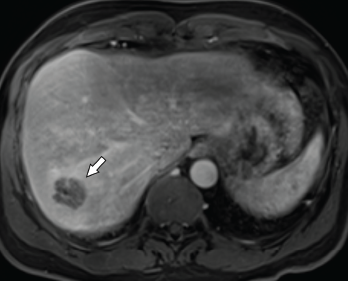
Figure 1. Axial CT of the liver, with the hepatic lobe abscess indicated by the arrow.

Abscesses are typically caused by infections, but some are, instead, sterile. Aseptic abscesses (AAs) are characterized by the same neutrophil-rich histo­pathology as infectious abscesses; however, they don’t improve with antibiotics. Rather, AAs require treatment with anti-inflammatory medications. Although relatively rare, this phenomenon is important for rheumatologists to recognize given its frequent association with under­lying systemic inflammatory diseases.

You Might Also Like
  • Fellows’ Forum Case Report: Autoinflammatory Diseases and Roles of Genetic, Molecular Testing
  • Case Report: A Patient Presents with Rare, Fulminant SAPHO Syndrome
  • Case Report: An Unusual Presentation of Neuro-Behçet’s Disease
Explore This Issue
June 2022

We describe two cases of AA seen by an inpatient rheumatology consultation service within a single year.

ad goes here:advert-1
ADVERTISEMENT
SCROLL TO CONTINUE

Case 1

A 46-year-old man presented to the hospital with fever and abdominal pain. His history was notable for six hospitalizations during the previous four years for recurrent hepatic abscesses. During each of these episodes, he had negative aerobic and anaerobic cultures of abscess aspirates and blood. Additional negative microbiological testing included human immunodeficiency virus (HIV), Coxiella, echinococcus and Entamoeba histolytica. Universal polymerase chain reaction testing (completed through the University of Washington Medical Center laboratory) for bacteria, fungi, mycobacterium tuberculous and nontuberculous mycobacterium was negative.

Axial CT showing a chest wall abscess.

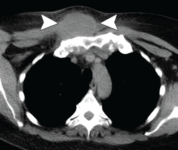
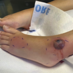
Figure 2. Axial CT showing a chest wall abscess.

His condition had temporarily improved with antibiotics during all but the last of the previous episodes. For that hospitalization, he was treated for a possible anatomic abnormality with placement of a biliary stent. He had experienced no recurrent abscess formation over the subsequent 18 months until the current hospitalization.

ad goes here:advert-2
ADVERTISEMENT
SCROLL TO CONTINUE

During the current hospitalization, the patient’s vital signs on admission were notable for a temperature of 103.5°F and sinus tachycardia of 110 beats per minute.

The initial laboratory testing demon­strated mild leukocytosis with a total white blood cell (WBC) count of 12.9×10⁹/L (reference range [RR]: 4.0–11.0×10⁹/L), and elevated inflammatory markers with an erythrocyte sedimentation rate (ESR) of 82 mm/hour (RR: 0–15 mm/hour for men younger than 50) and a C-reactive protein (CRP) of 32.6 mg/dL (RR: 0–0.6 mg/dL).

Imaging revealed four, right-lobe, hepatic abscesses measuring up to 3.0 cm in diameter (see Figure 1, above left), as well as an anorectal abscess and possible fistula of the gluteal fold.

ad goes here:advert-3
ADVERTISEMENT
SCROLL TO CONTINUE

One should have a strong suspicion for aseptic abscesses in a patient with abscesses that do not appropriately respond to antibiotic therapy.

Broad-spectrum antibiotics were initiated.

Axial CT showing a second chest wall abscess.

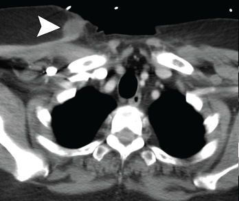
Figure 3. Axial CT showing a second chest wall abscess.

Aerobic and anaerobic cultures of abscess aspirate failed to identify an organism. Fungal and acid-fast bacillus (AFB) cultures were negative. Additional tests, including hepatitis B and C serologies, anti-Bartonella antibody, Fungitell, blastomyces antigen and cryptococcal antigen, were all negative.

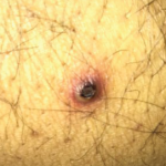
The patient’s history was also notable for recurrent, oral, aphthous ulcers and pyoderma gangrenosum in the preceding year. Due to the concern for occult inflammatory bowel disease (IBD), fecal calprotectin was obtained, but was indeterminate at 89.7 mg/kg (negative: <50 mg/kg).

Colonoscopy demonstrated a possible perianal fistula and a single rectal ulcer. Biopsy of the terminal ileum was unremark­able. Rectal ulcer biopsy demonstrated non-specific reactive changes, and a biopsy of perianal tissue demonstrated acute and chronic inflammation with ulcer and abscess. X-ray and magnetic resonance imaging (MRI) of the pelvis demonstrated sacroiliitis, despite a lack of inflammatory back pain.

Due to a lack of improvement with antimicrobials and a high suspicion for IBD, antibiotics were stopped, and he was started on 40 mg of prednisone daily. His fever and abdominal pain resolved. Adalimumab was initiated with no recurrence of abscesses to the present time.

Case 2

Pages: 1 2 3 4 | Single Page

Filed Under: Conditions Tagged With: abscess, aseptic abscess, case report, SAPHOIssue: June 2022

You Might Also Like:
  • Fellows’ Forum Case Report: Autoinflammatory Diseases and Roles of Genetic, Molecular Testing
  • Case Report: A Patient Presents with Rare, Fulminant SAPHO Syndrome
  • Case Report: An Unusual Presentation of Neuro-Behçet’s Disease
  • Case Report: An Adult-Onset Still’s Disease Mimic

Simple Tasks

Learn more about the ACR’s public awareness campaign and how you can get involved. Help increase visibility of rheumatic diseases and decrease the number of people left untreated.

Visit the Simple Tasks site »

Meeting Abstracts

Browse and search abstracts from the ACR Convergence and ACR/ARP Annual Meetings going back to 2012.

Visit the Abstracts site »

Rheumatology Research Foundation

The Foundation is the largest private funding source for rheumatology research and training in the U.S.

Learn more »

The Rheumatologist newsmagazine reports on issues and trends in the management and treatment of rheumatic diseases. The Rheumatologist reaches 11,500 rheumatologists, internists, orthopedic surgeons, nurse practitioners, physician assistants, nurses, and other healthcare professionals who practice, research, or teach in the field of rheumatology.

About Us / Contact Us / Advertise / Privacy Policy / Terms of Use / Cookie Preferences

  • Connect with us:
  • Facebook
  • Twitter
  • LinkedIn
  • YouTube
  • Feed

Copyright © 2006–2023 American College of Rheumatology. All rights reserved.

ISSN 1931-3268 (print)
ISSN 1931-3209 (online)