Video: Every Case Tells a Story| Webinar: ACR/CHEST ILD Guidelines in Practice

An official publication of the ACR and the ARP serving rheumatologists and rheumatology professionals

  • Conditions
    • Axial Spondyloarthritis
    • Gout and Crystalline Arthritis
    • Myositis
    • Osteoarthritis and Bone Disorders
    • Pain Syndromes
    • Pediatric Conditions
    • Psoriatic Arthritis
    • Rheumatoid Arthritis
    • Sjögren’s Disease
    • Systemic Lupus Erythematosus
    • Systemic Sclerosis
    • Vasculitis
    • Other Rheumatic Conditions
  • FocusRheum
    • ANCA-Associated Vasculitis
    • Axial Spondyloarthritis
    • Gout
    • Psoriatic Arthritis
    • Rheumatoid Arthritis
    • Systemic Lupus Erythematosus
  • Guidance
    • Clinical Criteria/Guidelines
    • Ethics
    • Legal Updates
    • Legislation & Advocacy
    • Meeting Reports
      • ACR Convergence
      • Other ACR meetings
      • EULAR/Other
    • Research Rheum
  • Drug Updates
    • Analgesics
    • Biologics/DMARDs
  • Practice Support
    • Billing/Coding
    • EMRs
    • Facility
    • Insurance
    • QA/QI
    • Technology
    • Workforce
  • Opinion
    • Patient Perspective
    • Profiles
    • Rheuminations
      • Video
    • Speak Out Rheum
  • Career
    • ACR ExamRheum
    • Awards
    • Career Development
  • ACR
    • ACR Home
    • ACR Convergence
    • ACR Guidelines
    • Journals
      • ACR Open Rheumatology
      • Arthritis & Rheumatology
      • Arthritis Care & Research
    • From the College
    • Events/CME
    • President’s Perspective
  • Search

Case Report: Atypical Presentation of Idiopathic Retroperitoneal Fibrosis

Roshniben Patel, MD, Simon Go, MD, Akhila Mohan, MD, & Maria Pardi, MD  |  Issue: March 2022  |  March 14, 2022

Figure 1

Figure 1

Retroperitoneal fibrosis (RPF) is a rare condition characterized by aberrant fibroinflammatory tissue developing in the retroperitoneum. This disorder was initially called Ormond’s disease. RPF may be idiopathic or secondary to other conditions. Idiopathic RPF is a part of the disease spectrum of chronic periaortitis due to its typical periaortoiliac localization.

Idiopathic retroperitoneal fibrosis is a relatively uncommon condition characterized by chronic fibroinflammatory proliferation in the retroperitoneum, usually around the infrarenal abdominal aorta and iliac vessels.1,2 It can lead to compression of retroperitoneal structures and a myriad of associated complications, most commonly obstructive uropathy and acute renal injury that can progress to end-stage renal disease.1,2,3

ad goes here:advert-1
ADVERTISEMENT
SCROLL TO CONTINUE

Due to its nonspecific and atypical clinical manifestations, prompt and early diagnosis can be challenging. Prognosis is mainly determined by the impact on renal function and complications from systemic corticosteroids.2

Case Presentation

A 40-year-old Black woman presented to the hospital complaining of nausea, decreased oral intake, diffuse abdominal pain and profuse watery diarrhea, all of which had persisted over a few weeks. She was found to have severe acute kidney injury with a serum creatinine of 9.3 mg/dL. Her baseline serum creatinine had been between 0.7 mg/dL and 0.8 mg/dL in prior years. She did not have a history of abdominal surgery or a family history of renal, oncologic or rheumatic conditions. She had never smoked cigarettes, although she had smoked cannabis for years. A urinalysis did not demonstrate evidence of an active sediment.

ad goes here:advert-2
ADVERTISEMENT
SCROLL TO CONTINUE

A non-contrast computed tomography (CT) scan of the abdomen and pelvis demonstrated mild symmetric prominence of the renal pelvises (see Figure 1, top), which was thought to be an incidental finding. A renal ultrasound showed mild bilateral hydronephrosis (see Figure 2, bottom) but the kidneys were found to be of normal size and echogenicity. An evaluation by a nephrologist and a gastroenterologist led to the conclusion that she likely had suffered severe gastrointestinal fluid losses, possibly due to gastroenteritis, which in turn led to acute tubular injury due to severe dehydration.

Renal ultrasound, sagittal view, showing mild bilateral hydronephrosis (arrows) with expansion of the renal sinus and calyces with normal renal cortical thickness and echogenicity.

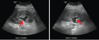
Figure 2. Renal ultrasound, sagittal view, showing mild bilateral hydronephrosis (arrows) with expansion of the renal sinus and calyces with normal renal cortical thickness and echogenicity.

The patient was treated with intravenous hydration and supportive care.  Her serum creatinine declined to 1.9 mg/dL, and her gastrointestinal symptoms had resolved by the time of discharge. During this hospitalization, she had also been diagnosed with right-sided Bell’s palsy, for which she received a one-week course of prednisone and valacyclovir. She was also diagnosed with hypertension, which was treated with amlodipine.

A month later, the patient was seen for follow-up by her primary care provider. Laboratory studies showed her serum creatinine level had increased to 5.96 mg/dL; she was asked to return to the hospital for further evaluation. At the time of this second admission, she noted two weeks of fatigue, generalized weakness and constant bilateral flank pain. She had intermittently loose stools but no recurrence of severe abdominal pain or diarrhea. She did not have urinary symptoms or hematuria, and she was not taking non-steroidal anti-inflammatory drugs or supplements.

The patient’s physical examination was significant for hypertension, tachycardia and non-pitting edema of the left leg. The abdomen was soft, without tenderness or guarding. There was no costovertebral angle tenderness.

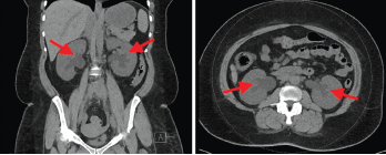
Laboratory findings demonstrated a blood urea nitrogen of 49 mg/dL and a serum creatinine of 11.1 mg/dL. Her urinalysis was unremarkable. A non-contrast CT scan of the abdomen and pelvis revealed a retroperitoneal density around the aortic bifurcation. Pelvic masses and mesenteric adenopathy were not seen (see Figure 3). A renal ultrasound (see Figure 4) showed grade 2 bilateral hydronephrosis. A lower extremity ultrasound did not demonstrate evidence of deep venous thrombosis.

Page: 1 2 3 4 | Single Page
Share: 

Filed under:Conditions Tagged with:case reportfibrosiskidneyOrmond's Diseaseretroperitoneal fibrosis (RPF)

Related Articles
    IgG4-Related Kidney Disease: Diagnostics, Manifestations, & More

    IgG4-Related Kidney Disease: Diagnostics, Manifestations & More

    May 17, 2018

    Immunoglobin G4-related disease (IgG4-RD) is a rare fibro-inflammatory disease of unknown etiology that has been recently recognized. It can cause fibro-inflammatory masses in almost every organ of the body and is associated with dense lymphoplasmacytic infiltration of IgG4-postitive plasma cells, storiform fibrosis and elevated levels of serum IgG4.1 IgG4-RD is a systemic disease that may…

    A Spotlight on IgG4-Related Disease

    January 1, 2013

    What rheumatologists need to know about identifying and diagnosing immunoglobulin G4-related disease (IgG4-RD)

    Case Report: Sarcoidosis in Patient with History of IgG4-Related Disease

    September 14, 2021

    Sarcoidosis and IgG4-related disease (IgG4-RD) are both immune-mediated, often multi-organ, diseases of uncertain etiology capable of presenting with diverse clinical manifestations. Many clinical features are common to both conditions, including hypergammaglobulinemia, the ability to form inflammatory masses and involvement of the lymph nodes, lacrimal glands, salivary glands, meninges and lungs. Although imaging modalities, such as…

    Fibrotic Diseases of the Retroperitoneum & Skin, & Rare Scleroderma Mimics

    January 19, 2018

    SAN DIEGO—Fibrosis affects all organ systems, but isn’t always systemic sclerosis. Experts on less common forms discussed patient presentations, diagnosis and treatment at the 2017 ACR/ARHP Annual Meeting in San Diego on Nov. 6. Retroperitoneal Fibrosis Formerly called Ormond’s disease, retroperitoneal fibrosis (RPF) is usually an IgG4-related disease, but has some unique characteristics, said John…

  • About Us
  • Meet the Editors
  • Issue Archives
  • Contribute
  • Advertise
  • Contact Us
  • Copyright © 2025 by John Wiley & Sons, Inc. All rights reserved, including rights for text and data mining and training of artificial technologies or similar technologies. ISSN 1931-3268 (print). ISSN 1931-3209 (online).
  • DEI Statement
  • Privacy Policy
  • Terms of Use
  • Cookie Preferences