Video: Every Case Tells a Story| Webinar: ACR/CHEST ILD Guidelines in Practice

An official publication of the ACR and the ARP serving rheumatologists and rheumatology professionals

  • Conditions
    • Axial Spondyloarthritis
    • Gout and Crystalline Arthritis
    • Myositis
    • Osteoarthritis and Bone Disorders
    • Pain Syndromes
    • Pediatric Conditions
    • Psoriatic Arthritis
    • Rheumatoid Arthritis
    • Sjögren’s Disease
    • Systemic Lupus Erythematosus
    • Systemic Sclerosis
    • Vasculitis
    • Other Rheumatic Conditions
  • FocusRheum
    • ANCA-Associated Vasculitis
    • Axial Spondyloarthritis
    • Gout
    • Psoriatic Arthritis
    • Rheumatoid Arthritis
    • Systemic Lupus Erythematosus
  • Guidance
    • Clinical Criteria/Guidelines
    • Ethics
    • Legal Updates
    • Legislation & Advocacy
    • Meeting Reports
      • ACR Convergence
      • Other ACR meetings
      • EULAR/Other
    • Research Rheum
  • Drug Updates
    • Analgesics
    • Biologics/DMARDs
  • Practice Support
    • Billing/Coding
    • EMRs
    • Facility
    • Insurance
    • QA/QI
    • Technology
    • Workforce
  • Opinion
    • Patient Perspective
    • Profiles
    • Rheuminations
      • Video
    • Speak Out Rheum
  • Career
    • ACR ExamRheum
    • Awards
    • Career Development
  • ACR
    • ACR Home
    • ACR Convergence
    • ACR Guidelines
    • Journals
      • ACR Open Rheumatology
      • Arthritis & Rheumatology
      • Arthritis Care & Research
    • From the College
    • Events/CME
    • President’s Perspective
  • Search

Case Report: Atypical Presentation of Idiopathic Retroperitoneal Fibrosis

Roshniben Patel, MD, Simon Go, MD, Akhila Mohan, MD, & Maria Pardi, MD  |  Issue: March 2022  |  March 14, 2022

Computed tomography (CT) scan of the abdomen and pelvis (left panel: coronal view; right panel: axial view), without intravenous contrast administration, demonstrating increased retroperitoneal density around the aortic bifurcation (arrows).

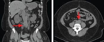
Figure 3. Computed tomography (CT) scan of the abdomen and pelvis (left panel: coronal view; right panel: axial view), without intravenous contrast administration, demonstrating increased retroperitoneal density around the aortic bifurcation (arrows).

The patient underwent retrograde pyelography, which confirmed bilateral hydronephrosis. A double-J stent was placed on the left kidney; because of the fishhook configuration of the right ureter, a nephroureteral stent was placed in the right kidney with subsequent conversion into a double-J stent.

Figure Renal ultrasound, sagittal view, showing bilateral grade 2 hydronephrosis (arrows) with normal renal cortical thickness and without focal lesions or nephrolithiasis.

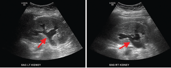
Figure 4. Renal ultrasound, sagittal view, showing bilateral grade 2 hydronephrosis (arrows) with normal renal cortical thickness and without focal lesions or nephrolithiasis.

A follow-up ultrasound demonstrated resolution of the obstruction (see Figure 5). Additional laboratory tests (see Table 1), including anti-nuclear antibody (ANA), anti-neutrophil cytoplasmic antibody (ANCA), complement, immunoglobulin levels, serum protein electrophoresis (SPEP) and immunofixation (IFE), were all unremarkable.

ad goes here:advert-1
ADVERTISEMENT
SCROLL TO CONTINUE

A clinical diagnosis of idiopathic retroperitoneal fibrosis leading to obstructive uropathy was made, and induction therapy with 1 mg/kg/day of prednisone was initiated. She responded well, and, at the time of discharge, her serum creatinine was 1.3 mg/dL; her improvement in serum creatinine was presumed to be due to stent placement and glucocorticoid therapy.

Renal ultrasound, sagittal view, performed a week after stenting showed complete resolution of hydronephrosis.

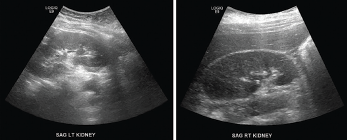
Figure 5. Renal ultrasound, sagittal view, performed a week after stenting showed complete resolution of hydronephrosis.

Discussion

RPF is a rare inflammatory disease often associated with non-specific clinical features; its etiopathogenesis is poorly understood.1 The overall incidence of RPF is unknown. Previous studies have estimated the incidence of idiopathic RPF at approximately 0.1–1.3 cases/100,000 people/year and the prevalence at approximately 1.4 cases/100,000 people.1 RPF is often diagnosed in patients between the ages of 40 and 60 years. Some studies have shown a 2:1 to 3:1 male predominance.1 It is possible that a higher number of male patients seek medical attention due to the genitourinary symptoms and, therefore, men may be over-represented in these studies.3

ad goes here:advert-2
ADVERTISEMENT
SCROLL TO CONTINUE

The age at the time of diagnosis and the male-to-female ratio have been changing over time. Comparing studies from 1987 and 2009, the age at the time of diagnosis increased from 56 to 64 years and male-to-female ratio increased to 3.3:1.4 Although some studies suggested a higher prevalence among white people than among other races, most of the studies noted no significant difference.

RPF is idiopathic in about two-thirds of cases; in one-third of cases, it is associated with other autoimmune diseases, such as IgG4-related disease (IgG4-RD).1 In this patient, the serum IgG4 level was normal; however, serum IgG4 is not a reliable indicator of IgG4-RD because a large proportion of patients with RPF associated with IgG4 RD can have normal IgG4 levels. On the other hand, patients with other inflammatory or proliferative conditions can have elevated levels of serum IgG4.5 Given these uncertainties, the authors cannot excluded the possibility that this patient had IgG4-RD instead of idiopathic RPF.

An association between RPF and other conditions, such as atherosclerotic aortic disease, drugs, infections (mainly bacterial and fungal), malignancy and radiation, has also been identified.3 In a retrospective cohort study of patients with RPF, it was noted that one patient had chronic hepatitis E infection, although causality could not be proved.6 No other data regarding the association between RPF and viral infections was found.

Environmental and genetic factors contribute to disease susceptibility. Tobacco smoke and asbestos exposure are strong risk factors for idiopathic RPF. Exposure to asbestos in a smoker is associated with doubling the risk of RPF.7 This susceptibility involves a CD4+ T cell-mediated immune response, which causes proliferation of B cells and fibroblasts in the periaortic retroperitoneum. The main genetic association is with HLA-DRB1*03, which has been associated with other autoimmune diseases.2

Local and systemic production of interleukin (IL) 6 and Th2 cytokines have been demonstrated in idiopathic RPF and RPF found in association with IgG4-RD.8 No data have been found linking cannabinoid use with the development of retroperitoneal fibrosis.

The most common manifestations of RPF include pain in the lower back, abdomen or flank and lower extremity edema due to inferior vena cava or lymphatic compression.1,3 More than 50% of male patients exhibit testicular symptoms, including testicular pain, with or without hydrocele, varicocele or both; these are thought to occur due to encasement of the spermatic vein, retrograde ejaculation and erectile dysfunction.3

Less common features include claudication due to compromise of the arterial circulation in the lower extremities and abdominal pain due to mesenteric ischemia.3 New-onset renovascular hypertension has been noted in 30–60% of patients. Laboratory findings may reveal acute kidney injury in 40–70% of patients. Elevated inflammatory markers, such as C-reactive protein and erythrocyte sedimentation rate, have been found in 50% to over 80% of cases.1,3 ANA are found in 60% of cases. Serum IL‑6 may also be elevated, reflecting an acute phase response; its correlation with disease activity or prognosis is unexplored.

In patients who present with acute kidney injury from obstruction, the diagnosis may be delayed due to lack of significant hydronephrosis on imaging, as demonstrated in this case. Compression of the kidneys, aorta and renal arteries by inflammatory and fibrotic tissue leads to decreased renal per­fusion and could cause hydrouretero­nephrosis.3 In patients with RPF, the degree of rise in serum creatinine is often out of proportion to the degree of hydronephrosis.

The diagnosis is made via imaging studies, such as computed tomography or magnetic resonance imaging. Retroperitoneal biopsy is only indicated in cases with atypical localization or with clinical findings suggestive for an alternative diagnosis.

Management

Management involves pharmacological as well as surgical approaches. The goal of idiopathic RPF treatment is to relieve ureteral obstruction via nephrostomy for patients with obstructive uropathy, followed by anti-fibrotic therapy to achieve disease regression.1 The cornerstone of medical therapy is corticosteroids, which suppress the synthesis of the cytokines involved in the fibroinflammatory response.9 Radio­graphic improvement is observed after about a week of treatment. Evaluation is recommended a month later to assess for disease response.9

Tamoxifen is commonly used as an alternative therapy if corticosteroids are contraindicated. In one small retrospective study, corticosteroids were found to be superior to tamoxifen for ameliorating symptoms, but patients who responded to tamoxifen had a lower chance of recurrence than those treated with corticosteroids.10

If remission is obtained, prednisone can be tapered to 5–10 mg/day and then maintained for an additional six to nine months.9 Studies are still ongoing regarding the use of biologics, such as rituximab and infliximab. Rituximab is now a standard therapy for the treatment of IgG4-RD.11 Other agents that can be used to induce remission include cyclophosphamide, mycophenolate mofetil and azathioprine.12

Table 1: Laboratory Test Results

LaboratoryPatientReference Range
IgG1,214 mg/dL700–1,600 mg/dL
IgG1629 mg/dL240–1,118 mg/dL
IgG2504 mg/dL124–549 mg/dL
IgG387 mg/dL21–134 mg/dL
IgG460 mg/dL1–123 mg/dL
IgA157 mg/dL68–408 mg/dL
IgM109 mg/dL35–264 mg/dL
Anti-nuclear antibody (ANA) screen immunofluorescence assayNegativeNegative
Anti-neutrophil cytoplasmic antibody (ANCA) IgG<1:20<1:20
Ganglioside (GM1) IgG4 IV0–50 IV
Ganglioside (GM1) IgM5 IV0–50 IV
Complement C3144 mg/dL90–180 mg/dL
Complement C440 mg/dL10–40 mg/dL
Antistreptolysin screen<55 IU/mL0–330 IU/mL
Serum protein electrophoresis (SPEP) and immunofixation (IFE)Slight restriction of protein migration in the gamma region. Normal IFE pattern, no monoclonal proteins.
Free κ light chains7.64 mg/dL0.33–1.94 mg/dL
Free λ light chains2.65 mg/dL0.57–2.63 mg/dL
Free κ/λ light chain ratio2.880.26–1.65

Page: 1 2 3 4 | Single Page
Share: 

Filed under:Conditions Tagged with:case reportfibrosiskidneyOrmond's Diseaseretroperitoneal fibrosis (RPF)

Related Articles
    IgG4-Related Kidney Disease: Diagnostics, Manifestations, & More

    IgG4-Related Kidney Disease: Diagnostics, Manifestations & More

    May 17, 2018

    Immunoglobin G4-related disease (IgG4-RD) is a rare fibro-inflammatory disease of unknown etiology that has been recently recognized. It can cause fibro-inflammatory masses in almost every organ of the body and is associated with dense lymphoplasmacytic infiltration of IgG4-postitive plasma cells, storiform fibrosis and elevated levels of serum IgG4.1 IgG4-RD is a systemic disease that may…

    A Spotlight on IgG4-Related Disease

    January 1, 2013

    What rheumatologists need to know about identifying and diagnosing immunoglobulin G4-related disease (IgG4-RD)

    Case Report: Sarcoidosis in Patient with History of IgG4-Related Disease

    September 14, 2021

    Sarcoidosis and IgG4-related disease (IgG4-RD) are both immune-mediated, often multi-organ, diseases of uncertain etiology capable of presenting with diverse clinical manifestations. Many clinical features are common to both conditions, including hypergammaglobulinemia, the ability to form inflammatory masses and involvement of the lymph nodes, lacrimal glands, salivary glands, meninges and lungs. Although imaging modalities, such as…

    Fibrotic Diseases of the Retroperitoneum & Skin, & Rare Scleroderma Mimics

    January 19, 2018

    SAN DIEGO—Fibrosis affects all organ systems, but isn’t always systemic sclerosis. Experts on less common forms discussed patient presentations, diagnosis and treatment at the 2017 ACR/ARHP Annual Meeting in San Diego on Nov. 6. Retroperitoneal Fibrosis Formerly called Ormond’s disease, retroperitoneal fibrosis (RPF) is usually an IgG4-related disease, but has some unique characteristics, said John…

  • About Us
  • Meet the Editors
  • Issue Archives
  • Contribute
  • Advertise
  • Contact Us
  • Copyright © 2025 by John Wiley & Sons, Inc. All rights reserved, including rights for text and data mining and training of artificial technologies or similar technologies. ISSN 1931-3268 (print). ISSN 1931-3209 (online).
  • DEI Statement
  • Privacy Policy
  • Terms of Use
  • Cookie Preferences