Video: Every Case Tells a Story| Webinar: ACR/CHEST ILD Guidelines in Practice

An official publication of the ACR and the ARP serving rheumatologists and rheumatology professionals

  • Conditions
    • Axial Spondyloarthritis
    • Gout and Crystalline Arthritis
    • Myositis
    • Osteoarthritis and Bone Disorders
    • Pain Syndromes
    • Pediatric Conditions
    • Psoriatic Arthritis
    • Rheumatoid Arthritis
    • Sjögren’s Disease
    • Systemic Lupus Erythematosus
    • Systemic Sclerosis
    • Vasculitis
    • Other Rheumatic Conditions
  • FocusRheum
    • ANCA-Associated Vasculitis
    • Axial Spondyloarthritis
    • Gout
    • Psoriatic Arthritis
    • Rheumatoid Arthritis
    • Systemic Lupus Erythematosus
  • Guidance
    • Clinical Criteria/Guidelines
    • Ethics
    • Legal Updates
    • Legislation & Advocacy
    • Meeting Reports
      • ACR Convergence
      • Other ACR meetings
      • EULAR/Other
    • Research Rheum
  • Drug Updates
    • Analgesics
    • Biologics/DMARDs
  • Practice Support
    • Billing/Coding
    • EMRs
    • Facility
    • Insurance
    • QA/QI
    • Technology
    • Workforce
  • Opinion
    • Patient Perspective
    • Profiles
    • Rheuminations
      • Video
    • Speak Out Rheum
  • Career
    • ACR ExamRheum
    • Awards
    • Career Development
  • ACR
    • ACR Home
    • ACR Convergence
    • ACR Guidelines
    • Journals
      • ACR Open Rheumatology
      • Arthritis & Rheumatology
      • Arthritis Care & Research
    • From the College
    • Events/CME
    • President’s Perspective
  • Search

Case Report: Coccidioides Immitis Infects a Patient’s Vascular Graft

Manjinder Kaur, DO, & Sabina Mian, MD  |  Issue: January 2019  |  January 17, 2019

Figures 1A (left) & 1B (right): A CT scan with abdominal and pelvic axial (a) and sagittal (b) views showing inflammatory changes (red arrow) adjacent to the aorta.

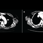
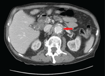
Figures 1A & 1B (below): A CT scan with abdominal and pelvic axial (a) and sagittal (b) views showing inflammatory changes (red arrow) adjacent to the aorta.

A 76-year-old Caucasian male with a history of abdominal aortic aneurysm repair five years earlier presented with three months duration of worsening periumbilical abdominal pain associated with nausea, non-bloody emesis, decreased appetite, fatigue and a 40 lb. weight loss. He denied having fever, chills, night sweats, temporal headaches, vision loss, chest pain, shortness of breath and jaw, upper and lower extremity pain.

On physical exam, the patient had no temporal artery tenderness. His peripheral pulses were normal and symmetric. The abdominal exam was significant for tenderness in the left periumbilical region with no guarding or rebound tenderness.

ad goes here:advert-1
ADVERTISEMENT
SCROLL TO CONTINUE

Laboratory analysis revealed a white blood cell count of 10.7 k/mm3, hemoglobin level of 11.1 g/dL and platelets at 294 k/mm3. The sedimentation rate of 140 mm/hr was elevated (normal: 0–22 mm/hr), as was the C-reactive protein level at 23.2 mg/L (normal: 2.0–8.0 mg/L). The creatinine level was normal at 0.93 mg/dL (normal: 0.6–1.50 mg/dL). The albumin level was low at 2.2 g/dL (normal: 3.4–5 g/dL).

ACase Report: Coccidioides Immitis Infects a Patient’s Vascular Graft 
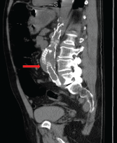
computed tomography (CT) scan of the abdomen and pelvis with contrast showed wall thickening and adjacent fat stranding involving the abdominal aorta and proximal bilateral common iliac arteries at the level of endovascular stent for the abdominal aortic aneurysm (see Figures 1a and 1b, right). These findings were suspicious for aortitis.

ad goes here:advert-2
ADVERTISEMENT
SCROLL TO CONTINUE

The patient was started on 40 mg intravenous methylprednisolone every eight hours. He experienced significant improvement in his abdominal pain and appetite.

A rheumatologist was consulted and recommended discontinuation of the methylprednisolone due to concern for infectious aortitis.

A vascular surgeon performed an aortic endograft explant and aortobiiliac bypass with CryoLife allograft. A large amount of pus surrounding the endograft was evident during surgery. The surgery was complicated by a splenic bleed requiring splenectomy. Histopathology revealed multiple necrotizing granulomas.

Aortic endograft cultures grew Coccidioides species. Serum Coccidioides complement fixation was positive 1:32 (normal <1:2) and (1-3)-β-d-glucan assay was elevated >500 pg/mL.

Initially, the patient was started on broad-spectrum anti-fungal coverage, with 375 mg isavuconazonium sulfate intravenously every eight hours. He received six doses. Then, he was switched to 375 mg isavuconazonium sulfate intravenously once daily with 5 mg/kg liposomal amphotericin B once daily. Once the mold was speciated to Coccidioides, the patient was taken off the isavuconazonium sulfate and switched to 400 mg intravenous fluconazole daily. The liposomal amphotericin B was continued for disseminated coccidioidomycosis.

The patient’s hospital course was complicated by retroperitoneal hematoma, prolonged ileus, hiatal hernia requiring exploratory laparotomy, acute renal failure requiring continuous renal replacement therapy, hemopneumothorax requiring chest tubes and acute respiratory failure requiring intubation. Unfortunately, his medical condition continued to deteriorate, and he died after transitioning to hospice care.

Page: 1 2 | Single Page
Share: 

Filed under:Conditions Tagged with:aortitis

Related Articles

    Fellow’s Forum Case Report: Takayasu’s Arteritis

    May 1, 2014

    How this rare form of large-vessel vasculitis affects different portions, branches of aorta and ways to diagnose, treat and manage the disease

    BYUNG H. BAN, DO

    Rheumatology Case Report: Immune-Related Aortitis Associated with Ipilimumab

    May 17, 2017

    Ipilimumab (Yervoy) is a monoclonal antibody directed against cytotoxic T-lymphocyte antigen 4 (CTLA-4). It was the first drug to demonstrate a survival benefit in advanced melanoma and was approved by the FDA in 2011.1 By blocking the CTLA-4 receptor, ipilimumab enhances the immune response against tumors via cytotoxic T lymphocyte activation and proliferation.2 However, immunopotentiating…

    Two Inflammatory Conditions—Polymyalgia Rheumatica and Giant Cell Arteritis—Share Clinical Connection

    March 1, 2013

    Polymyalgia rheumatica (PMR) and giant cell arteritis (GCA) have common clinical and epidemiologic links, but they need not occur synchronously

    Vasculitis

    June 1, 2009

    A New perspective on when and how it begins

  • About Us
  • Meet the Editors
  • Issue Archives
  • Contribute
  • Advertise
  • Contact Us
  • Copyright © 2025 by John Wiley & Sons, Inc. All rights reserved, including rights for text and data mining and training of artificial technologies or similar technologies. ISSN 1931-3268 (print). ISSN 1931-3209 (online).
  • DEI Statement
  • Privacy Policy
  • Terms of Use
  • Cookie Preferences