Video: Every Case Tells a Story| Webinar: ACR/CHEST ILD Guidelines in Practice

An official publication of the ACR and the ARP serving rheumatologists and rheumatology professionals

  • Conditions
    • Axial Spondyloarthritis
    • Gout and Crystalline Arthritis
    • Myositis
    • Osteoarthritis and Bone Disorders
    • Pain Syndromes
    • Pediatric Conditions
    • Psoriatic Arthritis
    • Rheumatoid Arthritis
    • Sjögren’s Disease
    • Systemic Lupus Erythematosus
    • Systemic Sclerosis
    • Vasculitis
    • Other Rheumatic Conditions
  • FocusRheum
    • ANCA-Associated Vasculitis
    • Axial Spondyloarthritis
    • Gout
    • Psoriatic Arthritis
    • Rheumatoid Arthritis
    • Systemic Lupus Erythematosus
  • Guidance
    • Clinical Criteria/Guidelines
    • Ethics
    • Legal Updates
    • Legislation & Advocacy
    • Meeting Reports
      • ACR Convergence
      • Other ACR meetings
      • EULAR/Other
    • Research Rheum
  • Drug Updates
    • Analgesics
    • Biologics/DMARDs
  • Practice Support
    • Billing/Coding
    • EMRs
    • Facility
    • Insurance
    • QA/QI
    • Technology
    • Workforce
  • Opinion
    • Patient Perspective
    • Profiles
    • Rheuminations
      • Video
    • Speak Out Rheum
  • Career
    • ACR ExamRheum
    • Awards
    • Career Development
  • ACR
    • ACR Home
    • ACR Convergence
    • ACR Guidelines
    • Journals
      • ACR Open Rheumatology
      • Arthritis & Rheumatology
      • Arthritis Care & Research
    • From the College
    • Events/CME
    • President’s Perspective
  • Search

Case Report: Elderly White Woman Presents with Recurrent Skin Lesions

Julia Tan, MD, Kun Huang, MD, PhD, Natasha Dehghan, MD, & Neda Amiri, MD   |  Issue: September 2019  |  September 17, 2019

These hematoxylin and eosin stain images show the patient’s histology of RCEV demonstrating vasculitis, including image A, a biopsy from her right leg showing ulceration and superficial and deep perivascular inflammation with vascular damage (magnification 40X); B, a biopsy from her left leg showing cutaneous small vessel vasculitis (the arrows indicate examples of damaged blood vessels) with fibrinoid necrosis of vessel walls and surrounding nuclear debris/leukocytoclasis (magnification 200X); and C, a biopsy from her right leg showing small vessel vasculitis (arrows), uninvolved adjacent small arterioles and abundant perivascular and interstitial eosinophils (magnification 200X).

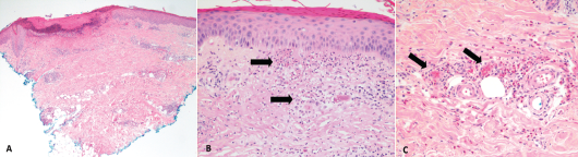
Figure 2. These hematoxylin and eosin stain images show the patient’s histology of RCEV demonstrating vasculitis, including image A, a biopsy from her right leg showing ulceration and superficial and deep perivascular inflammation with vascular damage (magnification 40X); B, a biopsy from her left leg showing cutaneous small vessel vasculitis (the arrows indicate examples of damaged blood vessels) with fibrinoid necrosis of vessel walls and surrounding nuclear debris/leukocytoclasis (magnification 200X); and C, a biopsy from her right leg showing small vessel vasculitis (arrows), uninvolved adjacent small arterioles and abundant perivascular and interstitial eosinophils (magnification 200X).

Discussion

RCEV was initially described by Chen et al. in 1994 and is characterized clinically by its benign course and classically pruritic skin eruptions. Various skin morphologies have been noted with RCEV, including urticaria, papules, patches, nodules, angioedema and vesicles.1-9 Only one previous group has described RCEV with ulcerating lesions, as we saw in this case.8 The lack of peripheral eosinophilia noted in our case was also unique, and this feature was noted in only three of the 17 cases.4,6,9

Histology can reveal necrotizing vasculitis with predominant eosinophilic infiltration, and others have commented on the presence of neutrophils and lymphocytes.5,8,9 Granulomas and leukocytoclastic features are typically absent in RCEV, although our case did feature some evidence of leukocytoclasis on histology.1-9 A number of specific findings enabled us to rule out similar conditions, such as eosinophilic granulomatous polyangiitis (EGPA), urticarial vasculitis, Wells syndrome and hypereosinophilic syndrome (HES).

ad goes here:advert-1
ADVERTISEMENT
SCROLL TO CONTINUE

Classically, patients with EGPA have asthma, peripheral eosinophilia and leukocytoclastic vasculitis with granulomas on biopsy.10 Urticarial vasculitis typically presents with skin lesions that resolve with pronounced central clearing and hyperpigmentation, rather than vesicles and ulcers.11 Leukocytoclasia is also a key histological feature in urticarial vasculitis.11,12

Patients with Wells syndrome tend to have peripheral eosinophilia and granulomas with no features of vasculitis on biopsy.13 Finally, HES was unlikely in light of the normal bone marrow biopsy and absence of eosinophilia or visceral involvement.14

ad goes here:advert-2
ADVERTISEMENT
SCROLL TO CONTINUE

Despite some atypical features, our patient’s clinical and histological presentation were consistent with RCEV. Glucocorticoids effectively manage RCEV, but recurrence is common after tapering the dose.1-9,15 Some recommend initiation of oral prednisone at 0.5 mg/kg/day and tapering it at two weeks to reach the minimum necessary dose to maintain remission.16

However, only a few known cases of RCEV report successful withdrawal of systemic steroids.5-7 Sakuma-Oyama et al. described RCEV in a 27-year-old female maintained solely on 300 mg of suplatast tosilate daily, a medication also used in eczema and asthma management known to suppress immunoglobulin E, interleukin 4 and interleukin 5.5 Tanglertsampan et al. successfully treated RCEV with 150 mg of indomethacin daily.8 Another group used dapsone in conjunction with glucocorticoids to manage two patients with RCEV.15 Use of other immunosuppressive agents in the treatment of RCEV was described in only one case, in which 2 mg of oral tacrolimus daily allowed prednisone to be tapered.7

In our case, azathioprine actually provided some steroid-sparing benefit, and her prednisone dose was tapered to 5 mg daily. But unfortunately, gastrointestinal side effects (e.g., nausea and diarrhea) prohibited azathioprine’s use. Methotrexate and hydroxychloroquine proved ineffective. Tacrolimus in addition to methotrexate caused kidney injury. Further therapy escalation to stronger immunosuppression, such as mycophenolate or cyclophosphamide, has not been reported and was limited in this case due to recurrent urinary tract infections.

Page: 1 2 3 4 | Single Page
Share: 

Filed under:Conditions Tagged with:case reportEosinophiliarecurrent cutaneous eosinophilic vasculitis (RCEV)

Related Articles
    Kamolrat/SHUTTERSTOCK.COM

    Eosinophilia: A Diagnostic Evaluation Guide for Rheumatologists

    June 15, 2015

    Clinical Vignette A 45-year-old woman with long-standing asthma and chronic sinusitis has new-onset peripheral neuropathy, arthralgias, fatigue, progressive dyspnea and a nonproductive cough. She has never smoked and has no environmental exposures. Her medications include an albuterol metered-dose inhaler (which she uses daily); an inhaled corticosteroid, montelukast; and ibuprofen (which she takes occasionally). She is…

    Eosinophilic Gastroenteritis & Eosinophilic Ascites Associated with RA

    June 17, 2019

    Eosinophilic gastroenteritis (EGE) is a rare condition caused by eosinophilic infiltration of the gastrointestinal (GI) tract. The condition is subdivided into the GI layers it affects: mucosal, muscular and subserosal.1 EGE usually presents with non-specific GI symptoms, such as impaired motility, intestinal obstruction and, rarely, ascites.2 Below, we report a rare case of EGE leading…

    The Latest on Eosinophilic Granulomatosis with Polyangiitis

    July 19, 2018

    The past five years have been busier than usual for the Churg-Strauss syndrome. It was renamed eosinophilic granulomatosis with polyangiitis (EGPA).1 Longitudinal cohorts totaling 484 patients—approximately as many as all previous series combined—were described.2,3 A proposal was advanced to remove and rename a subset in which vasculitis may not be present.4 And shortly after the…

    This chest CT shows new left upper lobe groundglass opacity.

    Case Report: Could Myocarditis + Shortness of Breath = EGPA?

    August 16, 2019

    Eosinophilic granulomatosis with polyangiitis (EGPA), also known as Churg-Strauss syndrome or allergic granulomatosis and angiitis, is a rare small- and medium-vessel vasculitis. This disease was first described by American pathologists Jacob Churg and Lotte Strauss in 1951.1 Although the vasculitis is often not apparent in the initial phases of the disease, EGPA can affect any…

  • About Us
  • Meet the Editors
  • Issue Archives
  • Contribute
  • Advertise
  • Contact Us
  • Copyright © 2025 by John Wiley & Sons, Inc. All rights reserved, including rights for text and data mining and training of artificial technologies or similar technologies. ISSN 1931-3268 (print). ISSN 1931-3209 (online).
  • DEI Statement
  • Privacy Policy
  • Terms of Use
  • Cookie Preferences