Video: Every Case Tells a Story| Webinar: ACR/CHEST ILD Guidelines in Practice

An official publication of the ACR and the ARP serving rheumatologists and rheumatology professionals

  • Conditions
    • Axial Spondyloarthritis
    • Gout and Crystalline Arthritis
    • Myositis
    • Osteoarthritis and Bone Disorders
    • Pain Syndromes
    • Pediatric Conditions
    • Psoriatic Arthritis
    • Rheumatoid Arthritis
    • Sjögren’s Disease
    • Systemic Lupus Erythematosus
    • Systemic Sclerosis
    • Vasculitis
    • Other Rheumatic Conditions
  • FocusRheum
    • ANCA-Associated Vasculitis
    • Axial Spondyloarthritis
    • Gout
    • Psoriatic Arthritis
    • Rheumatoid Arthritis
    • Systemic Lupus Erythematosus
  • Guidance
    • Clinical Criteria/Guidelines
    • Ethics
    • Legal Updates
    • Legislation & Advocacy
    • Meeting Reports
      • ACR Convergence
      • Other ACR meetings
      • EULAR/Other
    • Research Rheum
  • Drug Updates
    • Analgesics
    • Biologics/DMARDs
  • Practice Support
    • Billing/Coding
    • EMRs
    • Facility
    • Insurance
    • QA/QI
    • Technology
    • Workforce
  • Opinion
    • Patient Perspective
    • Profiles
    • Rheuminations
      • Video
    • Speak Out Rheum
  • Career
    • ACR ExamRheum
    • Awards
    • Career Development
  • ACR
    • ACR Home
    • ACR Convergence
    • ACR Guidelines
    • Journals
      • ACR Open Rheumatology
      • Arthritis & Rheumatology
      • Arthritis Care & Research
    • From the College
    • Events/CME
    • President’s Perspective
  • Search

Case Report: GPA or IgG4-Related Disease in an 11-Year-Old Girl?

Geoffrey E. Thiele, MD, & Sara Haro, MD  |  Issue: April 2023  |  April 7, 2023

Sinus disease was also found within the right maxillary sinus, with complete obliteration and underlying periosteal thickening. Circumferential mucosal thickening, with aerated secretions within the left maxillary sinus extending into the left ethmoid infundibulum, was seen.

Due to these findings, the patient was admitted to the hospital for expedited evaluation by, and consultation with, both an otolaryngologist and an ophthalmologist.

ad goes here:advert-1
ADVERTISEMENT
SCROLL TO CONTINUE

Hospital Course

On admission—two months after her first emergency department visit—her physical examination showed proptosis of the right eye with slight edema, compared with the left eye. This was a new finding. Despite the proptosis and mild edema, extraocular movements were intact. She did have lymphadenopathy that was worse along the right side of her neck. Otherwise, the examination was unremarkable. During questioning, she said she began to have blurry vision while in the emergency department that day.

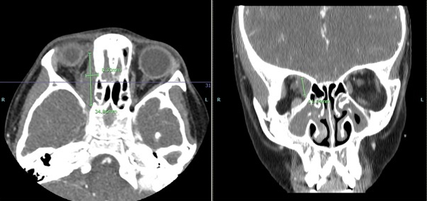
FIGURE 1. Click to enlarge.

Magnetic resonance imaging (MRI) showed a right medial, orbital mass encasing the right trochlear and medial rectus muscles (see Figure 1). The mass was non-uniform, with variable signal intensity. Again, imaging favored a non-infectious inflammatory process over infection. Pansinusitis with osseous destruction of the medial maxillary sinus walls was visualized on the right. The laboratory testing was expanded to include IgG subclasses to evaluate for IgG4 disease; all IgG subclasses were elevated (see Table 1).

ad goes here:advert-2
ADVERTISEMENT
SCROLL TO CONTINUE

The patient underwent biopsies of both the sinuses and the orbit shortly after admission. The hematoxylin and eosin (H&E) stain from the sinus biopsy showed densely collagenous fibrotic background with focal chronic inflamAnmation including many plasma cells in three of five fragments. Small vessels, including arterioles, had concentric or periarterial fibrosis with edematous changes. Focal neutrophilic vasculitis was noted. Classic fibrinoid necrosis was not seen on the slides examined. No granulomas, fungi or viral inclusions were identified. No evidence of neoplasm or malignancy was seen.

The biopsy from the orbit showed dense fibrous and adipose tissue with focal, chronic inflammation. Findings were difficult to fully examine due to extensive crush artifact (i.e., damaged tissue caused by iatrogenic compression of the tissue during collection; it can often result from firm handling with forceps, fingers or other instruments). Despite this, the small vessels were revealed to have concentric or periarterial fibrosis with edematous change. No granulomas, fungi or viral inclusions were identified, and no evidence of neoplasm or malignancy was seen.

Both biopsies were reviewed with the pathologist, and the findings from both the sinus and ocular biopsies appeared to be part of the same underlying process and not two different etiologies. A diagnosis of IgG4-related disease was considered unlikely based on her immunohistochemistry and laboratory testing (see Table 1). Given the biopsy results, early ANCA-associated vasculitis was considered more likely. Based on her presentation, biopsies and laboratory values, the patient was subsequently diagnosed with GPA.

Page: 1 2 3 4 5 | Single Page
Share: 

Filed under:ConditionsOther Rheumatic ConditionsVasculitis Tagged with:AAV FocusRheumcase reportFellowsFellows ForumGPAgranulomatosis with polyangiitisIgG4 related disease

Related Articles
    IgG4-Related Kidney Disease: Diagnostics, Manifestations, & More

    IgG4-Related Kidney Disease: Diagnostics, Manifestations & More

    May 17, 2018

    Immunoglobin G4-related disease (IgG4-RD) is a rare fibro-inflammatory disease of unknown etiology that has been recently recognized. It can cause fibro-inflammatory masses in almost every organ of the body and is associated with dense lymphoplasmacytic infiltration of IgG4-postitive plasma cells, storiform fibrosis and elevated levels of serum IgG4.1 IgG4-RD is a systemic disease that may…

    A Spotlight on IgG4-Related Disease

    January 1, 2013

    What rheumatologists need to know about identifying and diagnosing immunoglobulin G4-related disease (IgG4-RD)

    Case Report: Sarcoidosis in Patient with History of IgG4-Related Disease

    September 14, 2021

    Sarcoidosis and IgG4-related disease (IgG4-RD) are both immune-mediated, often multi-organ, diseases of uncertain etiology capable of presenting with diverse clinical manifestations. Many clinical features are common to both conditions, including hypergammaglobulinemia, the ability to form inflammatory masses and involvement of the lymph nodes, lacrimal glands, salivary glands, meninges and lungs. Although imaging modalities, such as…

    Figure 2: Renal Biopsy

    The Classification & Diagnosis of Granulomatosis with Polyangiitis

    August 16, 2018

    Based on the classification system developed by the Chapel Hill Consensus Conference, anti-neutrophil cytoplasmic antibody (ANCA) associated vasculitis is defined as a necrotizing vasculitis involving small vessels that is associated with myeloperoxidase (MPO) ANCA or proteinase 3 (PR3) ANCA and displays minimal immune deposits. The mechanism behind the pathogenesis of ANCA-associated vasculitis is not fully…

  • About Us
  • Meet the Editors
  • Issue Archives
  • Contribute
  • Advertise
  • Contact Us
  • Copyright © 2025 by John Wiley & Sons, Inc. All rights reserved, including rights for text and data mining and training of artificial technologies or similar technologies. ISSN 1931-3268 (print). ISSN 1931-3209 (online).
  • DEI Statement
  • Privacy Policy
  • Terms of Use
  • Cookie Preferences