Video: Every Case Tells a Story| Webinar: ACR/CHEST ILD Guidelines in Practice

An official publication of the ACR and the ARP serving rheumatologists and rheumatology professionals

  • Conditions
    • Axial Spondyloarthritis
    • Gout and Crystalline Arthritis
    • Myositis
    • Osteoarthritis and Bone Disorders
    • Pain Syndromes
    • Pediatric Conditions
    • Psoriatic Arthritis
    • Rheumatoid Arthritis
    • Sjögren’s Disease
    • Systemic Lupus Erythematosus
    • Systemic Sclerosis
    • Vasculitis
    • Other Rheumatic Conditions
  • FocusRheum
    • ANCA-Associated Vasculitis
    • Axial Spondyloarthritis
    • Gout
    • Psoriatic Arthritis
    • Rheumatoid Arthritis
    • Systemic Lupus Erythematosus
  • Guidance
    • Clinical Criteria/Guidelines
    • Ethics
    • Legal Updates
    • Legislation & Advocacy
    • Meeting Reports
      • ACR Convergence
      • Other ACR meetings
      • EULAR/Other
    • Research Rheum
  • Drug Updates
    • Analgesics
    • Biologics/DMARDs
  • Practice Support
    • Billing/Coding
    • EMRs
    • Facility
    • Insurance
    • QA/QI
    • Technology
    • Workforce
  • Opinion
    • Patient Perspective
    • Profiles
    • Rheuminations
      • Video
    • Speak Out Rheum
  • Career
    • ACR ExamRheum
    • Awards
    • Career Development
  • ACR
    • ACR Home
    • ACR Convergence
    • ACR Guidelines
    • Journals
      • ACR Open Rheumatology
      • Arthritis & Rheumatology
      • Arthritis Care & Research
    • From the College
    • Events/CME
    • President’s Perspective
  • Search

Case Report: GPA or IgG4-Related Disease in an 11-Year-Old Girl?

Geoffrey E. Thiele, MD, & Sara Haro, MD  |  Issue: April 2023  |  April 7, 2023

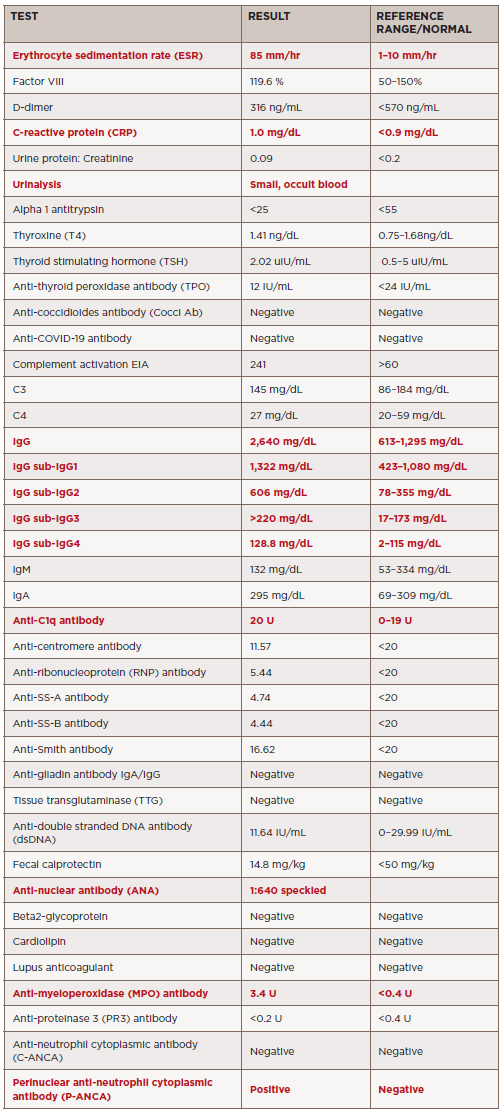
TABLE 1. Summary of laboratory results. Abnormal results indicated in red.

After diagnosis, the patient received pulse therapy with glucocorticoids (30 mg/kg/dose = 800 mg) for three days and then transitioned to 20 mg of oral prednisone twice a day, followed by a prednisone wean. She was given rituximab induction therapy (750 mg x two doses, two weeks apart) followed by maintenance therapy (thus far receiving one dose). Currently, she has been weaned off prednisone and is doing well overall, with minimal symptoms.

A follow-up MRI eight months later showed markedly decreased orbital mass, with only a small residual area of enhancement between the right medial rectus and superior oblique muscle. There was also slightly decreased pansinusitis, with osseous destruction of the medial maxillary sinus walls and dehiscence of the right lamina papyracea. Further, the anterior cranial fossa pachymeningeal thickening and enhancement was nearly resolved.

ad goes here:advert-1
ADVERTISEMENT
SCROLL TO CONTINUE

Discussion

Orbital lesions can be caused by a wide range of conditions, including infection, malignancy, foreign bodies, idiopathic or even autoimmune/inflammatory conditions. Those that are caused by inflammatory manifestations are rare in children, but some reports suggest an average onset at about 12 years of age.3,4 In a case series of children with orbital masses, the most common final diagnosis was granulomatosis with polyangiitis.3

Ocular findings in GPA are rarer in children than adults, where they can occur in up to 60% of cases. Reports indicate that up to 16% of adults initially present with orbital inflammatory involvement.5 Orbital GPA can be secondary to extension of sinus disease or isolated inflammation of the orbit itself.6 The most common presenting signs of orbital disease are ptosis, proptosis, pain, blurred vision, eyelid swelling and reduction of eye mobility.

ad goes here:advert-2
ADVERTISEMENT
SCROLL TO CONTINUE

A few case reports have discussed orbital tumors as initial manifestations of GPA in children.7 In one case series, four children presented with unilateral proptosis and ptosis due to an orbital tumor. Further, cases of GPA with orbital mass that can appear like IgG4-related disease have been reported.8 These presentations are strikingly similar to our patient. The tissues from our patient and the case report mentioned above both showed fibrosis with small and medium vessel vasculitis.

Although many of the clinical and radiographic findings are nonspecific for GPA, biopsy in conjunction with these findings can help differentiate this diagnosis from other potential etiologies, including IgG4-related disease. One general histologic finding that may be helpful in suggesting a diagnosis of GPA over other orbital inflammatory diseases is the presence of many scattered eosinophils, particularly eosinophils within granulomas.9

Page: 1 2 3 4 5 | Single Page
Share: 

Filed under:ConditionsOther Rheumatic ConditionsVasculitis Tagged with:AAV FocusRheumcase reportFellowsFellows ForumGPAgranulomatosis with polyangiitisIgG4 related disease

Related Articles
    IgG4-Related Kidney Disease: Diagnostics, Manifestations, & More

    IgG4-Related Kidney Disease: Diagnostics, Manifestations & More

    May 17, 2018

    Immunoglobin G4-related disease (IgG4-RD) is a rare fibro-inflammatory disease of unknown etiology that has been recently recognized. It can cause fibro-inflammatory masses in almost every organ of the body and is associated with dense lymphoplasmacytic infiltration of IgG4-postitive plasma cells, storiform fibrosis and elevated levels of serum IgG4.1 IgG4-RD is a systemic disease that may…

    A Spotlight on IgG4-Related Disease

    January 1, 2013

    What rheumatologists need to know about identifying and diagnosing immunoglobulin G4-related disease (IgG4-RD)

    Case Report: Sarcoidosis in Patient with History of IgG4-Related Disease

    September 14, 2021

    Sarcoidosis and IgG4-related disease (IgG4-RD) are both immune-mediated, often multi-organ, diseases of uncertain etiology capable of presenting with diverse clinical manifestations. Many clinical features are common to both conditions, including hypergammaglobulinemia, the ability to form inflammatory masses and involvement of the lymph nodes, lacrimal glands, salivary glands, meninges and lungs. Although imaging modalities, such as…

    Figure 2: Renal Biopsy

    The Classification & Diagnosis of Granulomatosis with Polyangiitis

    August 16, 2018

    Based on the classification system developed by the Chapel Hill Consensus Conference, anti-neutrophil cytoplasmic antibody (ANCA) associated vasculitis is defined as a necrotizing vasculitis involving small vessels that is associated with myeloperoxidase (MPO) ANCA or proteinase 3 (PR3) ANCA and displays minimal immune deposits. The mechanism behind the pathogenesis of ANCA-associated vasculitis is not fully…

  • About Us
  • Meet the Editors
  • Issue Archives
  • Contribute
  • Advertise
  • Contact Us
  • Copyright © 2025 by John Wiley & Sons, Inc. All rights reserved, including rights for text and data mining and training of artificial technologies or similar technologies. ISSN 1931-3268 (print). ISSN 1931-3209 (online).
  • DEI Statement
  • Privacy Policy
  • Terms of Use
  • Cookie Preferences