Video: Every Case Tells a Story| Webinar: ACR/CHEST ILD Guidelines in Practice

An official publication of the ACR and the ARP serving rheumatologists and rheumatology professionals

  • Conditions
    • Axial Spondyloarthritis
    • Gout and Crystalline Arthritis
    • Myositis
    • Osteoarthritis and Bone Disorders
    • Pain Syndromes
    • Pediatric Conditions
    • Psoriatic Arthritis
    • Rheumatoid Arthritis
    • Sjögren’s Disease
    • Systemic Lupus Erythematosus
    • Systemic Sclerosis
    • Vasculitis
    • Other Rheumatic Conditions
  • FocusRheum
    • ANCA-Associated Vasculitis
    • Axial Spondyloarthritis
    • Gout
    • Psoriatic Arthritis
    • Rheumatoid Arthritis
    • Systemic Lupus Erythematosus
  • Guidance
    • Clinical Criteria/Guidelines
    • Ethics
    • Legal Updates
    • Legislation & Advocacy
    • Meeting Reports
      • ACR Convergence
      • Other ACR meetings
      • EULAR/Other
    • Research Rheum
  • Drug Updates
    • Analgesics
    • Biologics/DMARDs
  • Practice Support
    • Billing/Coding
    • EMRs
    • Facility
    • Insurance
    • QA/QI
    • Technology
    • Workforce
  • Opinion
    • Patient Perspective
    • Profiles
    • Rheuminations
      • Video
    • Speak Out Rheum
  • Career
    • ACR ExamRheum
    • Awards
    • Career Development
  • ACR
    • ACR Home
    • ACR Convergence
    • ACR Guidelines
    • Journals
      • ACR Open Rheumatology
      • Arthritis & Rheumatology
      • Arthritis Care & Research
    • From the College
    • Events/CME
    • President’s Perspective
  • Search

Case Report: Is It Cardiac Sarcoidosis or COVID-19 Myocarditis?

Simranjit Kaur, MBBS, Sirajum Munira, MD, Farooq H. Sheikh, MD, FACC, & Anjani Pillarisetty, MD, RhMSUS  |  Issue: March 2023  |  March 12, 2023

A follow-up cardiac PET scan six months later showed complete resolution of active cardiac sarcoidosis, with improvement in EF to 40–45% on echocardiogram. Prednisone was weaned.

The patient developed a COVID-19 infection a few months later that did not require hospitalization.

ad goes here:advert-1
ADVERTISEMENT
SCROLL TO CONTINUE

A surveillance cardiac PET/CT scan four months after the infection showed interval development of metabolic activity within the basal anterior wall, mid to basal anterolateral wall, basal lateral and inferolateral regions, suggesting active cardiac sarcoidosis without evidence of extracardiac sarcoidosis. An echocardiogram showed worsening EF of 30–35%, with regional wall motion abnormalities and severely hypokinetic basal segments of the anterior and anterolateral walls.

He was restarted on prednisone taper, and mycophenolate was increased from 2 g daily to 3 g daily. His follow-up PET/CT scan showed no evidence of cardiac or extracardiac

ad goes here:advert-2
ADVERTISEMENT
SCROLL TO CONTINUE

sarcoidosis, and an echocardiogram showed improved EF of 40–45%.

Discussion

Mechanisms of COVID-19 mediated myocarditis include direct viral injury, systemic inflammation causing cytokine storm, stress-induced cardiomyopathy and microvascular thrombosis that can be focal or diffuse.1,2,5 Myocardial injury is most often seen later in the disease course, sometimes occurring several days after initial symptoms.

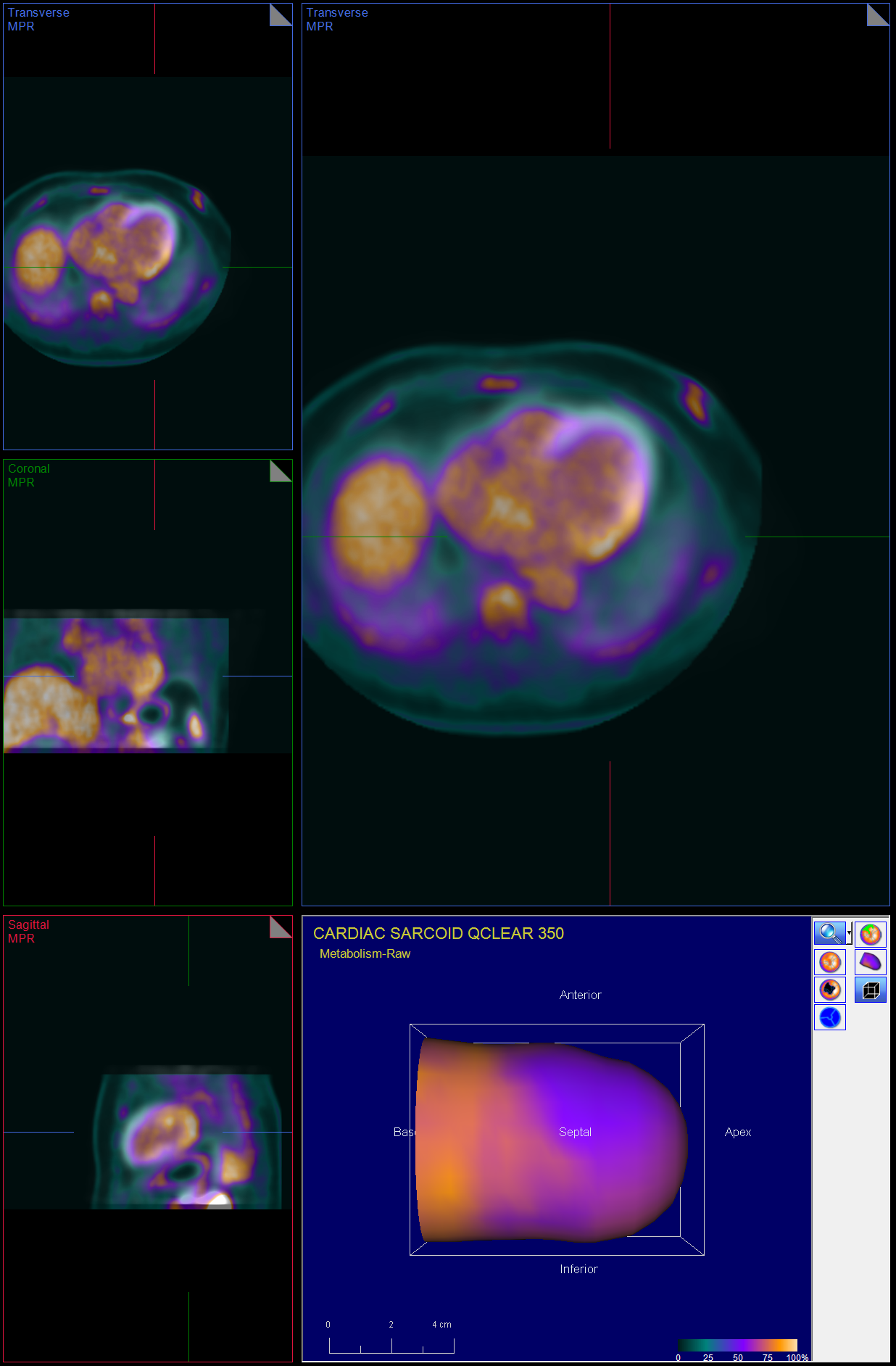
This image shows increased cardiac 18FDG uptake on the PET/CT nuclear scan without evidence of extracardiac inflammatory changes. (Click to enlarge.)

Even those who have clinically recovered from COVID-19 can have subclinical myocarditis. Depending on the extent of myocardial damage and time course of the disease, cardiac MRI abnormalities include diffuse myocardial edema, seen as regional or global hyperintensity; hyperemia, as evidenced by early gadolinium enhancement; and fibrosis, seen as a foci of late gadolinium enhancement.4-6 These findings depend on the extent of myocardial damage and time course of the infection. Similar imaging findings are seen in cardiac sarcoidosis, which has a high rate of recurrence with prednisone tapering.

It remains unclear whether the myocardial uptake seen on the PET/CT scan in our case was secondary to COVID-19 myocarditis vs. cardiac sarcoidosis, or whether COVID-19 plays a role in inducing the inflammatory process of sarcoidosis, although given the similar distribution of involvement with this patient’s prior cardiac sarcoidosis flares, one of the latter two scenarios seems more likely. Data on COVID-19 in patients with cardiac sarcoidosis are scarce, and further research is required to better understand the cardiac sequalae of this infection, as well as the effects of immunosuppressive therapies in these patients (see Figures 1 and 2).3

Page: 1 2 3 | Single Page
Share: 

Filed under:ConditionsOther Rheumatic Conditions Tagged with:case reportCOVID-19myocarditispulmonary sarcoidosisSarcoidosis

Related Articles

    Myositis & the Heart: New Perspectives on the Pathogenesis & Management of Cardiac Involvement in Myositis

    February 18, 2021

    Experts discuss the diagnosis and treatment of myositis-related cardiovascular disease.

    Sarcoidosis in the Spotlight: Screening, Treatment & More Insights into Sarcoidosis

    June 1, 2021

    An expert discussed the screening and treatment of sarcoidosis and drug-induced sarcoidosis-like reactions during the 2021 ACR State-of-the-Art Clinical Symposium.

    The Heart-SCC Puzzle

    May 1, 2009

    Identifying and treating cardiac involvement in systemic sclerosis can be a challenge

    Case Report: Sarcoidosis in Patient with History of IgG4-Related Disease

    September 14, 2021

    Sarcoidosis and IgG4-related disease (IgG4-RD) are both immune-mediated, often multi-organ, diseases of uncertain etiology capable of presenting with diverse clinical manifestations. Many clinical features are common to both conditions, including hypergammaglobulinemia, the ability to form inflammatory masses and involvement of the lymph nodes, lacrimal glands, salivary glands, meninges and lungs. Although imaging modalities, such as…

  • About Us
  • Meet the Editors
  • Issue Archives
  • Contribute
  • Advertise
  • Contact Us
  • Copyright © 2025 by John Wiley & Sons, Inc. All rights reserved, including rights for text and data mining and training of artificial technologies or similar technologies. ISSN 1931-3268 (print). ISSN 1931-3209 (online).
  • DEI Statement
  • Privacy Policy
  • Terms of Use
  • Cookie Preferences