Video: Every Case Tells a Story| Webinar: ACR/CHEST ILD Guidelines in Practice

An official publication of the ACR and the ARP serving rheumatologists and rheumatology professionals

  • Conditions
    • Axial Spondyloarthritis
    • Gout and Crystalline Arthritis
    • Myositis
    • Osteoarthritis and Bone Disorders
    • Pain Syndromes
    • Pediatric Conditions
    • Psoriatic Arthritis
    • Rheumatoid Arthritis
    • Sjögren’s Disease
    • Systemic Lupus Erythematosus
    • Systemic Sclerosis
    • Vasculitis
    • Other Rheumatic Conditions
  • FocusRheum
    • ANCA-Associated Vasculitis
    • Axial Spondyloarthritis
    • Gout
    • Psoriatic Arthritis
    • Rheumatoid Arthritis
    • Systemic Lupus Erythematosus
  • Guidance
    • Clinical Criteria/Guidelines
    • Ethics
    • Legal Updates
    • Legislation & Advocacy
    • Meeting Reports
      • ACR Convergence
      • Other ACR meetings
      • EULAR/Other
    • Research Rheum
  • Drug Updates
    • Analgesics
    • Biologics/DMARDs
  • Practice Support
    • Billing/Coding
    • EMRs
    • Facility
    • Insurance
    • QA/QI
    • Technology
    • Workforce
  • Opinion
    • Patient Perspective
    • Profiles
    • Rheuminations
      • Video
    • Speak Out Rheum
  • Career
    • ACR ExamRheum
    • Awards
    • Career Development
  • ACR
    • ACR Home
    • ACR Convergence
    • ACR Guidelines
    • Journals
      • ACR Open Rheumatology
      • Arthritis & Rheumatology
      • Arthritis Care & Research
    • From the College
    • Events/CME
    • President’s Perspective
  • Search

Case Report: Pneumatosis Cystoides Intestinalis

Jagmohan S. Jandu, MD, MA, Sri Harsha Boppana, MD, Prahlad A. Reddy, MD, & Atigadda N. Reddy, MD, FACP, FACG, AGAF  |  Issue: November 2023  |  November 8, 2023

Pneumatosis cystoides intestinalis (PCI) is a rare gastrointestinal pathological process defined by the presence of gas within the layers of the intestinal wall, commonly within the mucosa and submucosa of the small and large intestines.2,3 PCI has been described in the literature in association with various connective tissue diseases, including scleroderma, mixed connective tissue disease, Sjögren’s disease and systemic lupus erythematosus.1,3 Here, we present a case of PCI in a patient with limited systemic sclerosis.

Clinical Presentation

A 69-year-old woman presented to the rheumatology clinic to establish care. She was diagnosed with limited systemic sclerosis in 2007 by a dermatologist based on sclerodactyly, Raynaud’s phenomenon and telangiectasias. She had never seen a rheumatologist before, nor had she required treatment with any disease-modifying anti-rheumatic drug (DMARD).

ad goes here:advert-1
ADVERTISEMENT
SCROLL TO CONTINUE

She reported a history of gastroesophageal reflux disease and was followed by a gastroenterologist. She denied dysphagia, abdominal pain and distension. She denied any cardiopulmonary symptoms and had no known history of interstitial lung disease or pulmonary hypertension.

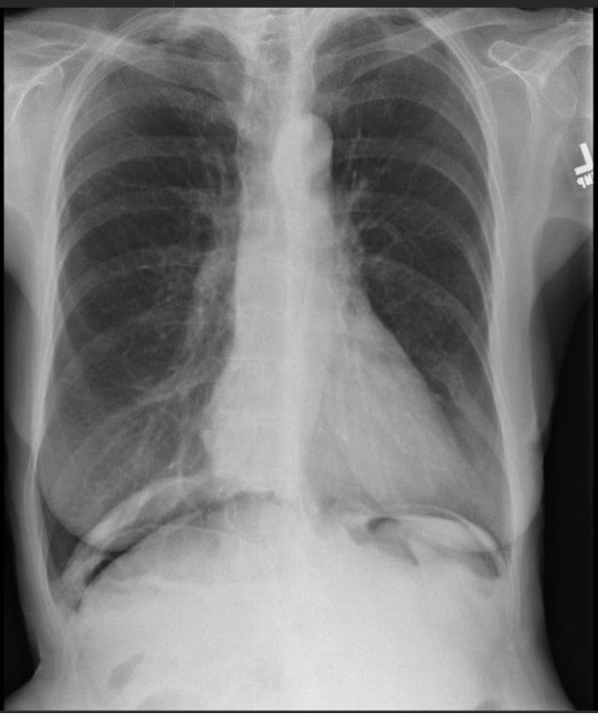
FIGURE 1: A chest X-ray incidentally revealed pneumoperitoneum. (Click to enlarge.)

Her medical history also included chronic musculoskeletal pain, for which she was treated chronically with ibuprofen and hydrocodone/acetaminophen. She had undergone a partial colectomy in 2011 and was unable to provide any specific reason or diagnosis for this procedure.

ad goes here:advert-2
ADVERTISEMENT
SCROLL TO CONTINUE

Physical examination revealed bilateral sclerodactyly distal to the proximal interphalangeal joints. Multiple telangiectasias were noted on the tongue and the dorsal and ventral aspects of both hands. The cardiac exam did not reveal a murmur. Pulmonary auscultation was notable for crackles at the base of the left lung. Diffuse soft tissue and joint tenderness were noted, without evidence of inflammatory arthritis.

Laboratory testing performed before her appointment revealed an anti-nuclear antibody titer of >1:1280 [reference range: negative] with a centromere pattern.

The patient’s abnormal lung exam was evaluated with a two-view chest X-ray. This incidentally revealed pneumoperitoneum (see Figure 1).1

Due to geographic and transportation constraints, the patient was unable to return to the clinic for a repeat evaluation; therefore, she was contacted by telephone. She reported having mild abdominal discomfort and bloating, the nature and duration of which she was not able to clearly describe. She denied any sudden severe abdominal pain, trauma, fever or recent abdominal surgery or procedures. Prompt evaluation at the nearest emergency department was recommended.

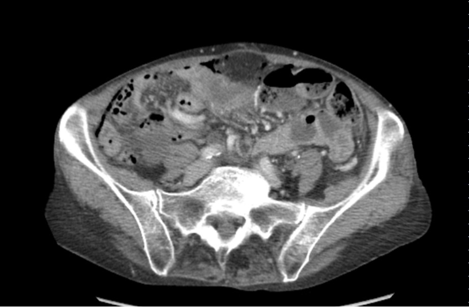
FIGURE 2: Computerized tomography of the abdomen with contrast confirmed pneumoperitoneum and revealed intramural air in the ileum without obvious gastrointestinal perforation. (Click to enlarge.)

Page: 1 2 3 | Single Page
Share: 

Filed under:ConditionsSystemic Sclerosis Tagged with:case reportpneumatosis cystoides intestinalis (PCI)

Related Articles

    Commensal Cross-Reactive T & B Cells Trigger Autoimmunity

    October 26, 2020

    Researchers are finding that antigens from the microbiota may play a role in the development of autoimmunity.

    okawa somchai / shutterstock.com

    Case Report: A Lupus Patient with Abdominal Pain

    February 16, 2021

    Our patient was a 33-year-old, 5’2″ Asian woman with a past medical history of systemic lupus erythematosus (SLE). The diagnosis was based on serologies positive for anti-nuclear antibodies (ANAs), as well as antibodies to Sm, RNP and SSA. Her illness included neuropsychiatric and cutaneous involvement. She also had a diagnosis of Hashimoto’s thyroiditis.  She presented…

    The Microbiome in Pediatric Rheumatic Diseases

    The Microbiome in Pediatric Rheumatic Diseases

    April 15, 2016

    The human intestinal microbiota is home to more than 1,000 bacterial species, containing approximately 3 million genes, many of which code for functions that have the potential to affect human physiology.1 Smaller numbers of organisms are also present in the skin, upper gastrointestinal tract, female reproductive tract and the oro- and nasopharynx. As tools have…

    Mattew - Bilder und mehr / shutterstock.com

    Yao Syndrome: A Case Report & Clinical Review

    November 12, 2020

    Case Presentation History of present illness A 66-year-old white woman presented with unexplained, recurrent episodes of high fever, abdominal pain, rash and arthralgias occurring over the previous three years. During typical episodes, the patient experienced flu-like symptoms, followed by fever, abdominal pain and non-bloody diarrhea without tenesmus. ad goes here:advert-1ADVERTISEMENTSCROLL TO CONTINUEHer temperatures were 101–103ºF,…

  • About Us
  • Meet the Editors
  • Issue Archives
  • Contribute
  • Advertise
  • Contact Us
  • Copyright © 2025 by John Wiley & Sons, Inc. All rights reserved, including rights for text and data mining and training of artificial technologies or similar technologies. ISSN 1931-3268 (print). ISSN 1931-3209 (online).
  • DEI Statement
  • Privacy Policy
  • Terms of Use
  • Cookie Preferences