Video: Every Case Tells a Story| Webinar: ACR/CHEST ILD Guidelines in Practice

An official publication of the ACR and the ARP serving rheumatologists and rheumatology professionals

  • Conditions
    • Axial Spondyloarthritis
    • Gout and Crystalline Arthritis
    • Myositis
    • Osteoarthritis and Bone Disorders
    • Pain Syndromes
    • Pediatric Conditions
    • Psoriatic Arthritis
    • Rheumatoid Arthritis
    • Sjögren’s Disease
    • Systemic Lupus Erythematosus
    • Systemic Sclerosis
    • Vasculitis
    • Other Rheumatic Conditions
  • FocusRheum
    • ANCA-Associated Vasculitis
    • Axial Spondyloarthritis
    • Gout
    • Psoriatic Arthritis
    • Rheumatoid Arthritis
    • Systemic Lupus Erythematosus
  • Guidance
    • Clinical Criteria/Guidelines
    • Ethics
    • Legal Updates
    • Legislation & Advocacy
    • Meeting Reports
      • ACR Convergence
      • Other ACR meetings
      • EULAR/Other
    • Research Rheum
  • Drug Updates
    • Analgesics
    • Biologics/DMARDs
  • Practice Support
    • Billing/Coding
    • EMRs
    • Facility
    • Insurance
    • QA/QI
    • Technology
    • Workforce
  • Opinion
    • Patient Perspective
    • Profiles
    • Rheuminations
      • Video
    • Speak Out Rheum
  • Career
    • ACR ExamRheum
    • Awards
    • Career Development
  • ACR
    • ACR Home
    • ACR Convergence
    • ACR Guidelines
    • Journals
      • ACR Open Rheumatology
      • Arthritis & Rheumatology
      • Arthritis Care & Research
    • From the College
    • Events/CME
    • President’s Perspective
  • Search

Case Report: Sternoclavicular Erosions in a Patient with Uncontrolled RA

Diana M. Girnita, MD, PhD, Avis Ware, MD, Sangita Kapur, MD, & Christine Chhakchhuak, MD  |  Issue: December 2015  |  December 17, 2015

Based on these symptoms, elevated disease activity score and evidence of new erosions at the level of SC joint and hands, abatacept was discontinued and rituximab was initiated. Two months later, his symptoms improved significantly and the DAS/ESR score decreased to 2.5.

It is well documented that the SC joint can be affected by traumatic or non-traumatic processes.

Discussion

The SC joint is an often neglected joint. Table 1 summarizes the pathology of the SC joint in the context of arthritides. This diarthrodial synovial joint can be affected in septic,5,14 rheumatoid,1,2,9,15 reactive,16 psoriatic11,17 and seronegative arthritis,7–9 including ankylosing spondylitis,18 gout,10 osteoarthrtitis6 or SAPHO syndrome (synovitis, acne, pustulosis, hyperostosis and osteitis).4,19
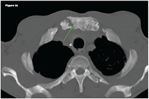
Figure 1A

Figure 1A

Dilsen et al reported that in patients with RA, the SC joint can be affected in 40% of patients.1 Involvement of this joint can cause chest pain, tenderness to palpation, pain in the shoulder area, hypertrophy and joint crepitus on the exam.1,6,7,9 Kalliomäki et al reported that 33 of 105 patients with RA had either subchondral or marginal erosions documented by tomography, whereas chest roentgenograms were usually unremarkable.15

ad goes here:advert-2
ADVERTISEMENT
SCROLL TO CONTINUE

In our patient, the SC changes were documented at the time of chest pain evaluation. However, in the context of his active disease, new erosions were interpreted as a marker of treatment failure to abatacept, suggesting that the disease remained poorly controlled. This prompted a repeat hand X-ray, which also showed new erosions. Based on these new findings, we decided to change his treatment to rituximab, and it was noted that the patient had significant improvement in his symptoms.
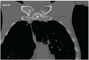
Figure 1B

Figure 1B
Non-contrast chest CT: Axial (Figure 1A) and coronal multiplanar reformat (Figure 1B) images demonstrate bilateral sternoclavicular joint erosive arthropathy, right more than left (green arrows). A joint effusion is not identified. Key: ST-sternum, C-clavicle

The SC joint is easily accessible and can be evaluated by ultrasound, CT or magnetic resonance.1,9,15,20,21 A recent ultrasound study evaluated the SC joint in 209 RA patients and compared the findings with a group of healthy controls.20 In the RA group, 19% of scanned joints were clinically involved (meaning that patients presented with pain or swelling of the joint) compared with only 4% in the control group. Further, 43% of RA patients evaluated with ultrasound had evidence of osteophytes (29%), synovitis (15%), erosions (11%) or intra-articular power Doppler (2%), suggesting synovial hyperperfusion characteristic of an inflammatory process. When compared with the group of healthy controls, all of these findings were statistically significant. The same study reported correlation between the presence of synovitis and intra-articular power Doppler, with a higher DAS28 score.20

Conclusion

Involvement of the SC joint is not uncommon in patients with active RA. It is easily accessible for evaluation via ultrasound or CT imaging.

ad goes here:advert-1
ADVERTISEMENT
SCROLL TO CONTINUE

Page: 1 2 3 4 | Single Page
Share: 

Filed under:ConditionsRheumatoid Arthritis Tagged with:ClinicalDiagnosisoutcomepatient careRARheumatoid arthritisSCsternoclavicular jointTreatment

Related Articles

    What Rheumatologists Should Look for in Diagnosing SAPHO Syndrome

    November 1, 2012

    SAPHO syndrome (synovitis, acne, pustulosis, hyperostosis, and osteitis) is an obscure condition that can vex patients and clinicians

    Musculoskeletal Ultrasound: A Valuable Tool for Diagnosing Rheumatic Illnesses

    Musculoskeletal Ultrasound: A Valuable Tool for Diagnosing Rheumatic Illnesses

    October 15, 2015

    Musculoskeletal (MSK) ultrasound is a valuable imaging modality for the practicing rheumatologist and provides an efficient tool with high diagnostic value in the evaluation of patients with musculoskeletal complaints. The use of MSK ultrasound has evolved in the U.S. due to the emergence of less-expensive, portable ultrasound units, which provide high-quality gray-scale and power Doppler…

    Tips for Implementing Ultrasound Training in Rheumatology Fellowships

    February 13, 2020

    ATLANTA—Point-of-care ultrasound education mainly has occurred at the undergraduate level at U.S. medical schools, but rheumatology fellowship training programs are rapidly catching up and integrating it into their curricula, according to two program directors who reviewed the state of rheumatology ultrasound education, including potential barriers to its implementation, on Nov. 12 at the 2019 ACR/ARP…

    Ultrasound Can be Useful in Diagnosing Gout

    June 14, 2017

    The presence of synovial monosodium urate monohydrate (MSU) crystals is the gold standard for diagnosing gout. But a new study, funded in part by the ACR and led by rheumatologists, including Alexis Ogdie, MD, MSCE, evaluated the effectiveness of ultrasound in diagnosing it. The study found that ultrasound can be useful in discriminating gout from non-gout….

  • About Us
  • Meet the Editors
  • Issue Archives
  • Contribute
  • Advertise
  • Contact Us
  • Copyright © 2025 by John Wiley & Sons, Inc. All rights reserved, including rights for text and data mining and training of artificial technologies or similar technologies. ISSN 1931-3268 (print). ISSN 1931-3209 (online).
  • DEI Statement
  • Privacy Policy
  • Terms of Use
  • Cookie Preferences