Video: Every Case Tells a Story| Webinar: ACR/CHEST ILD Guidelines in Practice

An official publication of the ACR and the ARP serving rheumatologists and rheumatology professionals

  • Conditions
    • Axial Spondyloarthritis
    • Gout and Crystalline Arthritis
    • Myositis
    • Osteoarthritis and Bone Disorders
    • Pain Syndromes
    • Pediatric Conditions
    • Psoriatic Arthritis
    • Rheumatoid Arthritis
    • Sjögren’s Disease
    • Systemic Lupus Erythematosus
    • Systemic Sclerosis
    • Vasculitis
    • Other Rheumatic Conditions
  • FocusRheum
    • ANCA-Associated Vasculitis
    • Axial Spondyloarthritis
    • Gout
    • Psoriatic Arthritis
    • Rheumatoid Arthritis
    • Systemic Lupus Erythematosus
  • Guidance
    • Clinical Criteria/Guidelines
    • Ethics
    • Legal Updates
    • Legislation & Advocacy
    • Meeting Reports
      • ACR Convergence
      • Other ACR meetings
      • EULAR/Other
    • Research Rheum
  • Drug Updates
    • Analgesics
    • Biologics/DMARDs
  • Practice Support
    • Billing/Coding
    • EMRs
    • Facility
    • Insurance
    • QA/QI
    • Technology
    • Workforce
  • Opinion
    • Patient Perspective
    • Profiles
    • Rheuminations
      • Video
    • Speak Out Rheum
  • Career
    • ACR ExamRheum
    • Awards
    • Career Development
  • ACR
    • ACR Home
    • ACR Convergence
    • ACR Guidelines
    • Journals
      • ACR Open Rheumatology
      • Arthritis & Rheumatology
      • Arthritis Care & Research
    • From the College
    • Events/CME
    • President’s Perspective
  • Search

Dermatology Symptoms Point to Connective Tissue Disorder

Natalie A. Wright, MD, and Joseph F. Merola, MD, MMSc, FAAD, FACR  |  Issue: May 2015  |  May 15, 2015

NotarYES/shutterstock.com

Image Credit: NotarYES/shutterstock.com

The Case

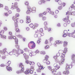
A 68-year-old woman with a past medical history of Charcot-Marie-Tooth presents with thickening of the skin on her trunk and extremities, which she has had for the past seven months (see Figures 1 and 2). Her symptoms first began with swelling of her bilateral upper and lower extremities. She is now having difficulty with writing and extending her legs. She feels well other­wise and has no other systemic complaints. On exam, she has induration of both forearms and the dorsal aspect of her hands and legs, extending from the feet to above the knees. She has flexion contractures of the fingers and knees (see Figure 3). Exam also reveals hyperpigmented, indurated plaques on the back, abdomen and inguinal folds (see Figure 4). Nailfold capillaroscopy is normal. There is no history of Raynaud’s. Laboratory findings include a normocytic anemia, ANA, Lyme titer and negative serum protein electrophoresis. The remainder of an extensive review of symptoms was unremarkable.

What’s Your Diagnosis?

  1. Systemic sclerosis
  2. Hypereosinophilic syndrome
  3. Lipodermatosclerosis
  4. Eosinophilic fasciitis
  5. Acrodermatitis chronica atrophicans

Correct Answer

D. Eosinophilic fasciitis

ad goes here:advert-1
ADVERTISEMENT
SCROLL TO CONTINUE

Eosinophilic fasciitis (EF) is a rare connective tissue disorder characterized by edema and induration of the extremities, and rarely the trunk, that can ultimately result in sclerosis and fibrosis, leading to joint contractures and disability. The cutaneous changes may be accompanied by peripheral eosinophilia, hypergammaglobulinemia, elevated acute-phase reactants and, rarely, underlying malignancy. Diagnosis and treatment can be challenging. Cutaneous clues can prevent misdiagnosis, which can lead to unnecessary tests, invasive workups and delays in treatment.

EF is usually a disease of middle-aged or older adults, but there are reports in children & young adults.

The pathogenesis of EF remains unknown; however, interleukin 5 and matrix metalloproteinase 13 have been postulated to play a role.1 Although strenuous exercise and trauma have been described as eliciting factors for EF, this preceding history tends to be reported in only 30–45% of patients.2 Several medications have been suggested as contributing factors in the development of EF, including statins, phenytoin, subcutaneous heparin and therapies for multiple sclerosis.3 Infectious agents, such as borrelia or mycoplasma, have been described as a possible cause in a minority of cases. Hematologic and myelodysplastic disorders, as well as a few reports of solid tumors, have been associated with EF, and treatment of the underlying disorder can result in resolution of the cutaneous findings.
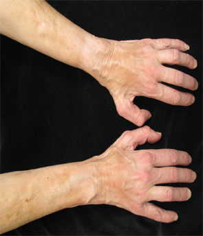
Figures 1 & 2: On exam, the patient had thickening of the skin on her extremities.

Figure 1.

ad goes here:advert-2
ADVERTISEMENT
SCROLL TO CONTINUE

Page: 1 2 3 4 | Single Page
Share: 

Filed under:ConditionsSystemic Sclerosis Tagged with:connective tissue disorderDermatologyeosinophilic fasciitis

Related Articles

    A Case of Eosinophilic Fasciitis Presenting with Pansclerotic Morphea

    May 17, 2019

    Eosinophilic fasciitis generally presents with the acute onset of edema followed by progressive skin induration in the setting of hypergammaglobulinemia, an elevated erythrocyte sedimentation rate (ESR) and peripheral eosinophilia in 63–93% of patients.1,2 Skin involvement is typically limited to 20.1% of total body surface area and most commonly involves the extremities symmetrically.1,3 The condition was…

    Fellows’ Forum Case Report: Palmar Fasciitis & Polyarthritis Syndrome

    November 16, 2015

    Case report: A 78-year-old Caucasian female presented to our outpatient rheumatology clinic with pain in her bilateral shoulders, hands and knees that began suddenly one month earlier. She admitted to stiffness in her hands lasting several hours, and expressed an inability to extend her fingers. She denied fever, rashes, jaw claudication, headache or visual changes….

    Kamolrat/SHUTTERSTOCK.COM

    Eosinophilia: A Diagnostic Evaluation Guide for Rheumatologists

    June 15, 2015

    Clinical Vignette A 45-year-old woman with long-standing asthma and chronic sinusitis has new-onset peripheral neuropathy, arthralgias, fatigue, progressive dyspnea and a nonproductive cough. She has never smoked and has no environmental exposures. Her medications include an albuterol metered-dose inhaler (which she uses daily); an inhaled corticosteroid, montelukast; and ibuprofen (which she takes occasionally). She is…

    Dermatology Case Answer

    February 3, 2012

    A 64-year-old man with history of type-II diabetes (well controlled on sitagliptin/metformin), hypertension, and dyslipidemia presents with complaints of an increasingly painful left lower-extremity lesion present for two to three months.

  • About Us
  • Meet the Editors
  • Issue Archives
  • Contribute
  • Advertise
  • Contact Us
  • Copyright © 2025 by John Wiley & Sons, Inc. All rights reserved, including rights for text and data mining and training of artificial technologies or similar technologies. ISSN 1931-3268 (print). ISSN 1931-3209 (online).
  • DEI Statement
  • Privacy Policy
  • Terms of Use
  • Cookie Preferences