Video: Every Case Tells a Story| Webinar: ACR/CHEST ILD Guidelines in Practice

An official publication of the ACR and the ARP serving rheumatologists and rheumatology professionals

  • Conditions
    • Axial Spondyloarthritis
    • Gout and Crystalline Arthritis
    • Myositis
    • Osteoarthritis and Bone Disorders
    • Pain Syndromes
    • Pediatric Conditions
    • Psoriatic Arthritis
    • Rheumatoid Arthritis
    • Sjögren’s Disease
    • Systemic Lupus Erythematosus
    • Systemic Sclerosis
    • Vasculitis
    • Other Rheumatic Conditions
  • FocusRheum
    • ANCA-Associated Vasculitis
    • Axial Spondyloarthritis
    • Gout
    • Psoriatic Arthritis
    • Rheumatoid Arthritis
    • Systemic Lupus Erythematosus
  • Guidance
    • Clinical Criteria/Guidelines
    • Ethics
    • Legal Updates
    • Legislation & Advocacy
    • Meeting Reports
      • ACR Convergence
      • Other ACR meetings
      • EULAR/Other
    • Research Rheum
  • Drug Updates
    • Analgesics
    • Biologics/DMARDs
  • Practice Support
    • Billing/Coding
    • EMRs
    • Facility
    • Insurance
    • QA/QI
    • Technology
    • Workforce
  • Opinion
    • Patient Perspective
    • Profiles
    • Rheuminations
      • Video
    • Speak Out Rheum
  • Career
    • ACR ExamRheum
    • Awards
    • Career Development
  • ACR
    • ACR Home
    • ACR Convergence
    • ACR Guidelines
    • Journals
      • ACR Open Rheumatology
      • Arthritis & Rheumatology
      • Arthritis Care & Research
    • From the College
    • Events/CME
    • President’s Perspective
  • Search

Diagnostic Challenges of SLE & Celiac Sprue

Leslie Pack Ranken, MD  |  Issue: March 2018  |  March 17, 2018

Renal biopsy showing class III lupus nephritis with rare foci of subendothelial deposits (wire loop lesions).

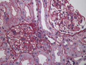
Figure 2. Renal biopsy showing class III lupus nephritis with rare foci of subendothelial deposits (wire loop lesions).

Due to his persistent pancytopenia, the consulting hematologist performed a bone marrow biopsy that did not demonstrate evidence of a malignancy. Within a week of his admission, his creatinine increased to 2.46 (mg/dL) from 0.98 (mg/dL) with a urinalysis that demonstrated mild hemoglobin and albumin. A renal biopsy was performed; it demonstrated evidence of lupus nephritis class III (see Figure 2). Approximately three weeks later, he was also found to have anti-double-stranded DNA antibodies.

The patient was started on prednisone 60  mg daily, hydroxychloroquine 400 mg daily and mycophenolate mofetil 2 g daily.

ad goes here:advert-1
ADVERTISEMENT
SCROLL TO CONTINUE

Two weeks later, the patient developed chest pain and was found to have a small pericardial effusion on echocardiogram, but a normal ejection fraction. That same day, he became confused, and a magnetic resonance imaging (MRI) of the brain was performed; it showed evidence of multiple emboli. Neurology and cardiology were consulted and felt the patient likely had Liebman-Sach’s endocarditis, although there was no lesion found on the valves. The mitral valve was thickened, however.

One day later, the patient developed third-degree atrioventricular block requiring a pacemaker. He was started on anticoagulation given the presence of multiple cerebral emboli. Because of his myo­carditis, which was thought to be due to lupus, and a new diagnosis of nephritis, he was switched from mycophenolate mofetil to cyclophosphamide, and all of his symptoms improved. He completed a six-month course of intravenous cyclophosphamide.

ad goes here:advert-2
ADVERTISEMENT
SCROLL TO CONTINUE

He is now taking a remission maintenance regimen of mycophenolate mofetil and hydroxychloroquine, and he remains on anticoagulation, given his history of embolic cerebrovascular events.

SLE is known to cause gastrointestinal symptoms & can occur in the form of … pancreatitis & even malabsorption.

Discussion

SLE is known to cause gastrointestinal symptoms and can occur in the form of serositis, vasculitis, pancreatitis and even malabsorption. Whether or not the malabsorption is from SLE itself or from a concurrent malabsorptive disease can be difficult to discern. Several case reports of concurrent celiac disease and SLE have appeared in the literature over the years. Celiac disease has been reported in other autoimmune diseases as well.

Our patient first presented with symptoms of malabsorption in the presence of an anti-TGA IgA antibody. Anti-TGA IgA antibody is 98–100% sensitive and specific for celiac disease when small bowel atrophy is present, as with our patient.1 However, his atrophy was mild, and the lymphocytes seen could be present in lupus alone. It is possible that the mild atrophy was due to mild malabsorption from SLE itself.

Page: 1 2 3 | Single Page
Share: 

Filed under:Systemic Lupus Erythematosus Tagged with:celiac diseaseceliac sprueLupusmalabsorptionSLE

Related Articles

    Celiac Awareness Makes a Difference for Rheumatology Patients

    September 24, 2018

    Celiac disease and gluten sensitivity are difficult to diagnose, but they affect multiple systems and are associated with other autoimmune diseases. Lan Chen, MD, PhD, believes better testing and clearer education will be a gamechanger for celiac patients…

    Insights into Celiac Disease & Systemic Autoimmune Diseases

    May 14, 2019

    Little is known about the immunopathogenic relationship between celiac disease and systemic autoimmune diseases, such as primary Sjögren’s syndrome and systemic sclerosis. But new research indicates that screening for celiac disease may benefit patients newly diagnosed with systemic autoimmune diseases, as well as supports the idea that celiac disease may accelerate the development of systemic autoimmune disease…

    Diagnostic Tests, Tips for Gluten-Induced Celiac Disease

    June 13, 2016

    CHICAGO—Celiac disease—the gluten-induced illness that can be seen alongside rheumatic diseases—has been seen much more commonly over the past 20 years than it was previously, but the illness can come with questions that are not always straightforward, an expert said at the ACR’s State-of-the-Art Clinical Symposium. The disease, in which the small intestine becomes inflamed…

    Celiac Disease in Pediatric Patients

    October 21, 2015

    A retrospective study shows that some patients presenting for pediatric rheumatology evaluation test positive for celiac disease—some without showing any gastrointestinal manifestations. Treatment includes a gluten-free diet…

  • About Us
  • Meet the Editors
  • Issue Archives
  • Contribute
  • Advertise
  • Contact Us
  • Copyright © 2025 by John Wiley & Sons, Inc. All rights reserved, including rights for text and data mining and training of artificial technologies or similar technologies. ISSN 1931-3268 (print). ISSN 1931-3209 (online).
  • DEI Statement
  • Privacy Policy
  • Terms of Use
  • Cookie Preferences