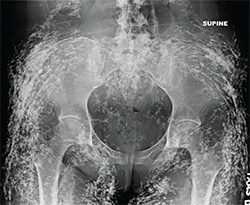
Figure 1:
Anteroposterior radiograph of the pelvis.
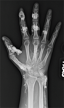
Figure 2: Anteroposterior radiograph of the right hand.
Figure 1:
Anteroposterior radiograph of the pelvis.
Figure 2: Anteroposterior radiograph of the right hand.
View the question. Findings/Diagnosis The radiographs demonstrate diffuse sheetlike and tumefactive calcifications throughout the subcutaneous tissues, muscle and fascia of the pelvis and right hand. ad goes here:advert-1ADVERTISEMENTSCROLL TO CONTINUEThe underlying bones and joint spaces appear normal. The differential diagnosis for soft tissue calcification is extensive and includes metabolic disturbances (particularly of calcium and phosphate),…
Radiograph, MR images reveal pigmented villonodular synovitis in 54-year-old woman with right elbow pain
Radiograph, MR images of 54-year-old woman with right elbow pain

Editor’s note: In this recurring feature, we first present a series of images (this page) for your review, and then a brief discussion of the findings and diagnosis. Before you turn to the discussion, examine these images carefully and draw your own conclusions. History A 33-year-old woman with a 16-year history of systemic lupus erythematosus…