Video: Every Case Tells a Story| Webinar: ACR/CHEST ILD Guidelines in Practice

An official publication of the ACR and the ARP serving rheumatologists and rheumatology professionals

  • Conditions
    • Axial Spondyloarthritis
    • Gout and Crystalline Arthritis
    • Myositis
    • Osteoarthritis and Bone Disorders
    • Pain Syndromes
    • Pediatric Conditions
    • Psoriatic Arthritis
    • Rheumatoid Arthritis
    • Sjögren’s Disease
    • Systemic Lupus Erythematosus
    • Systemic Sclerosis
    • Vasculitis
    • Other Rheumatic Conditions
  • FocusRheum
    • ANCA-Associated Vasculitis
    • Axial Spondyloarthritis
    • Gout
    • Psoriatic Arthritis
    • Rheumatoid Arthritis
    • Systemic Lupus Erythematosus
  • Guidance
    • Clinical Criteria/Guidelines
    • Ethics
    • Legal Updates
    • Legislation & Advocacy
    • Meeting Reports
      • ACR Convergence
      • Other ACR meetings
      • EULAR/Other
    • Research Rheum
  • Drug Updates
    • Analgesics
    • Biologics/DMARDs
  • Practice Support
    • Billing/Coding
    • EMRs
    • Facility
    • Insurance
    • QA/QI
    • Technology
    • Workforce
  • Opinion
    • Patient Perspective
    • Profiles
    • Rheuminations
      • Video
    • Speak Out Rheum
  • Career
    • ACR ExamRheum
    • Awards
    • Career Development
  • ACR
    • ACR Home
    • ACR Convergence
    • ACR Guidelines
    • Journals
      • ACR Open Rheumatology
      • Arthritis & Rheumatology
      • Arthritis Care & Research
    • From the College
    • Events/CME
    • President’s Perspective
  • Search

How to Diagnose Upper Extremity Injuries

Mike Fillon  |  Issue: February 2020  |  February 13, 2020

New Africa / shutterstock.com

New Africa / shutterstock.com

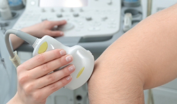
ATLANTA—Two ways to investigate injuries to the upper extremities are by in-depth physical examinations and ultrasound. In a Clinical Practice session at the 2019 ACR/ARP Annual Meeting, Anatomy: Correlating Physical Exam and Ultrasound in Common Sports Injuries of the Upper Extremity, Carlin Senter, MD, FACP, associate professor of primary care sports medicine at the University of California, San Francisco, and Anthony M. Reginato, MD, PhD, associate professor and director of the Division of Rheumatology at Brown University, Providence, R.I., presented strategies to diagnose different injuries via case studies.

Most sports medicine problems can be identified by conducting a careful history and physical exam and knowing your anatomy, said Dr. Senter. “However, musculoskeletal ultrasound is playing an increasingly important role in both diagnosis and guiding treatment for sports medicine problems.”

ad goes here:advert-1
ADVERTISEMENT
SCROLL TO CONTINUE

In the session, Dr. Reginato reviewed basic principles of ultrasound. An ultrasound transducer converts electrical energy into acoustic pulses (i.e., sound) and transmits them into the body. Through the piezoelectric effect (generation of an electric charge in response to applied mechanical stress) the transducer acts as a receiver of refracted echoes and converts them into electrical signals, which are finally processed as an image.

Going Gray

The content of various tissues will differentially reflect the transmitted pulses, resulting in different gray images. Hyperechoic structures (white on the screen) are those with an increased density of sound waves compared with surrounding structures, such as bone, tendon, nerves and calcification. Hypoechoic structures (gray on the screen), including the synovium, lymph nodes and some tumors, give off fewer echoes. Anechoic structures, appearing black on the screen, are those without an echo, such as fluid and cartilage.

ad goes here:advert-2
ADVERTISEMENT
SCROLL TO CONTINUE

‘Musculoskeletal ultrasound is playing an increasingly important role in both diagnosis & guiding treatment for sports medicine problems.’  —Carlin Senter, MD, FACP

“In rheumatology, ultrasound can predict diagnosis—as well as provide early detection—staging of the disease and monitor response to therapy,” said Dr. Reginato.

A Case of Shoulder Pain

In one case, a 57-year-old man presented with right shoulder pain that started after he had slipped and fallen three months earlier. He tried physical therapy without benefit and was waking up at night due to pain. On physical exam, the shoulder was not tender. The active range of motion of the shoulder was intact, with pain on abduction between 60º and 120º. He had moderate pain with resisted external rotation of the shoulder and was unable to perform a lift-off test. The diagnosis was a rotator cuff tendon tear.

Page: 1 2 3 | Single Page
Share: 

Filed under:ConditionsMeeting Reports Tagged with:2019 ACR/ARP Annual Meetingdiagnostic imagingphysical examsports medicineUltrasoundupper extremity injuries

Related Articles

    Using Ultrasound to Diagnose Carpal Tunnel Syndrome

    April 26, 2018

    Note: Updated May 2, 2018, to correct a link in the reference section. The error was introduced in editing. A 44-year-old Caucasian woman presented to the outpatient rheumatology clinic that had followed her for several years for rheumatoid arthritis. She was compliant with her regimen of hydroxychloroquine, etanercept and salsalate. Her chief complaint was worsening…

    How to Evaluate Shoulder Pain

    February 2, 2013

    The common causes of shoulder pain and diagnostic tests that rheumatologists need to know

    Musculoskeletal Ultrasound: A Valuable Tool for Diagnosing Rheumatic Illnesses

    Musculoskeletal Ultrasound: A Valuable Tool for Diagnosing Rheumatic Illnesses

    October 15, 2015

    Musculoskeletal (MSK) ultrasound is a valuable imaging modality for the practicing rheumatologist and provides an efficient tool with high diagnostic value in the evaluation of patients with musculoskeletal complaints. The use of MSK ultrasound has evolved in the U.S. due to the emergence of less-expensive, portable ultrasound units, which provide high-quality gray-scale and power Doppler…

    A transverse view of the ulnar groove in full elbow extension. The red arrow indicates the advancing edge of the MHTr.

    Recurrent Medial Elbow Pain Following Successful Tommy John Surgery

    August 12, 2020

    A 27-year-old, left-handed man was referred to our ultrasound clinic for left elbow pain. History The patient had been a pitcher on a Minor League Baseball team. Two years before, he developed sudden, severe medial elbow pain while pitching in a game. The pain was associated with some tingling down the left medial forearm. The…

  • About Us
  • Meet the Editors
  • Issue Archives
  • Contribute
  • Advertise
  • Contact Us
  • Copyright © 2025 by John Wiley & Sons, Inc. All rights reserved, including rights for text and data mining and training of artificial technologies or similar technologies. ISSN 1931-3268 (print). ISSN 1931-3209 (online).
  • DEI Statement
  • Privacy Policy
  • Terms of Use
  • Cookie Preferences