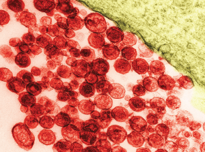
Particles of human T-lymphotropic virus 1 on a transmission electron micrograph. The virus has been implicated in lymphoma development.
David M. Phillips / Science Source
A 57-year-old Ghanaian woman was referred to our rheumatology practice with acute, left elbow swelling and pain. The referring oncologist suspected gout, because the patient had hyperuricemia.
Six months before, the patient was diagnosed with stage IV human T-lymphotropic virus type 1 (HTLV-1)-associated adult T cell lymphoma (ATLL). Her initial oncologic manifestations included multiple thoracic, abdominal and retroperitoneal masses. Biopsy of an abdominal mass showed histological and immunohistochemical features of T cell lymphoma with CD3 positivity. Serologic assay and Western blot were positive for HTLV-1. The patient had osteoblastic lesions of the skull, and axial and appendicular skeleton, including bilateral distal humerus diaphyses. She developed a pathologic fracture of the right mid-humerus that was treated conservatively.

