Video: Every Case Tells a Story| Webinar: ACR/CHEST ILD Guidelines in Practice

An official publication of the ACR and the ARP serving rheumatologists and rheumatology professionals

  • Conditions
    • Axial Spondyloarthritis
    • Gout and Crystalline Arthritis
    • Myositis
    • Osteoarthritis and Bone Disorders
    • Pain Syndromes
    • Pediatric Conditions
    • Psoriatic Arthritis
    • Rheumatoid Arthritis
    • Sjögren’s Disease
    • Systemic Lupus Erythematosus
    • Systemic Sclerosis
    • Vasculitis
    • Other Rheumatic Conditions
  • FocusRheum
    • ANCA-Associated Vasculitis
    • Axial Spondyloarthritis
    • Gout
    • Psoriatic Arthritis
    • Rheumatoid Arthritis
    • Systemic Lupus Erythematosus
  • Guidance
    • Clinical Criteria/Guidelines
    • Ethics
    • Legal Updates
    • Legislation & Advocacy
    • Meeting Reports
      • ACR Convergence
      • Other ACR meetings
      • EULAR/Other
    • Research Rheum
  • Drug Updates
    • Analgesics
    • Biologics/DMARDs
  • Practice Support
    • Billing/Coding
    • EMRs
    • Facility
    • Insurance
    • QA/QI
    • Technology
    • Workforce
  • Opinion
    • Patient Perspective
    • Profiles
    • Rheuminations
      • Video
    • Speak Out Rheum
  • Career
    • ACR ExamRheum
    • Awards
    • Career Development
  • ACR
    • ACR Home
    • ACR Convergence
    • ACR Guidelines
    • Journals
      • ACR Open Rheumatology
      • Arthritis & Rheumatology
      • Arthritis Care & Research
    • From the College
    • Events/CME
    • President’s Perspective
  • Search

IgG4-Related Disease: The Latest On Its Presentation, Diagnosis & Management

Jason Liebowitz, MD, FACR  |  Issue: May 2021  |  May 13, 2021

KATERYNA KON / Science Source

Pembrolizumab antibody, a humanized IgG4 antibody.
KATERYNA KON / Science Source

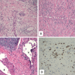
In 1888, Dr. Jan Mikulicz-Radecki reported a case of chronic, bilateral, painless enlargement of the salivary and lacrimal glands that appeared to be idiopathic.1 In subsequent years, other patients with these findings were reported, and the term Mikulicz syndrome was used to describe these cases. Although Mikulicz syndrome is now known to be associated with certain specific conditions, such as sarcoidosis and Sjögren’s syndrome, IgG4-related disease has also been identified as a potential cause.

At the 2021 ACR State-of-the-Art Clinical Symposium, John Stone, MD, MPH, professor of medicine and Edward A. Fox Chair in Medicine, Massachusetts General Hospital, Boston, provided an excellent update regarding the clinical presentation, diagnosis and management of IgG4-related disease.

ad goes here:advert-1
ADVERTISEMENT
SCROLL TO CONTINUE

The Condition

IgG4-related disease is a progressive, immune-mediated fibrotic disease that typically results in tumor-like masses in many affected organs, such as salivary and lacrimal glands, the pancreas and the kidneys. Studies have shown that CD4+ cytotoxic T lymphocytes are the predominant immune cell infiltrate in IgG4-related disease, with M2 macrophages, activated B cells and fibroblasts likely also playing a role in the generation of inflammatory masses. Classic histologic findings seen on biopsies of affected organs include lymphoplasmacytic infiltrate, obliterative phlebitis and storiform fibrosis, with a large proportion of the plasma cells staining positive for IgG4.2

Serum IgG4 concentration is a good biomarker for many patients with this condition, Dr. Stone noted. However, an elevated serum IgG4 level on its own does not clinch the diagnosis. In cases of IgG4-related disease, a high baseline serum IgG4 level can be helpful for following the disease longitudinally, because the level will often decrease with effective treatment or waning disease activity. If a patient is being treated for their IgG4-related disease and the serum IgG4 level begins to rise, clinicians should consider changing the treatment regimen.

ad goes here:advert-2
ADVERTISEMENT
SCROLL TO CONTINUE

Other biomarkers of disease may include dissociation between erythrocyte sedimentation rate (ESR) and C-reactive protein (CRP)—with high ESR and low CRP being a typical finding in active disease—elevated IgG1 and serum IgE levels, low C3 and C4 levels, and peripheral eosinophilia.

A major advancement in the study of this condition arrived in the form of the 2019 ACR/European League Against Rheumatism (EULAR) classification criteria for IgG4-related disease.3 These criteria employ a three-step process to accurately identify patients with this disease and seek to exclude mimicking conditions.

Step 1: Identify typical involvement in at least one of the 11 possible organs typically affected by IgG4-related disease (i.e., pancreas, salivary glands, bile ducts, orbits, lacrimal glands, kidneys, lungs, aorta, retro­peritoneum, pachymeninges or thyroid gland [Riedel’s thyroiditis]). Such involvement can be based on clinical or radiographic assessment, or pathologic evidence of an inflammatory process accompanied by a lymphoplasmacytic infiltrate of uncertain etiology in one of these organs.

Step 2: Apply exclusion criteria, which includes a total of 32 clinical, serologic, radiographic and pathologic items. This large array of exclusion criteria is meant to ensure patients with a wide range of autoimmune, hematologic, infectious and malignant conditions are not misdiagnosed as having IgG4-related disease. Example: Leukopenia and thrombocytopenia are unusual in IgG4-related disease, but often found in myelodysplastic syndromes, hematopoietic cancers and connective tissue diseases, such as systemic lupus erythematosus. Similarly, certain radiographic findings, such as long-bone abnormalities seen in Erdheim-Chester disease, should not be attributed to IgG4-related disease.

Step 3: Use eight weighted inclusion criteria domains, which include immuno­staining of IgG positive cells, head and neck gland involvement, pancreatic and biliary tree involvement, renal involvement, chest involvement and findings of retroperitoneal fibrosis or periaortitis. In studying these classification criteria in several validation cohorts, a threshold of 20 points on the criteria scale has a sensitivity of 97–99% and a specificity of 82–85%.3

Dr. Stone explained that both a proliferative and fibrotic subtype of IgG4-related disease appear to exist. In the proliferative subtype, patients commonly experience multi-organ involvement, and typical lab findings include elevated IgG4, IgG1 and IgE levels, eosinophilia and hypocomplementemia.

Responsiveness to treatment is typically quite good in these patients. In individuals with the fibrotic subtype, patients often experience involvement of the retroperitoneum, aorta and periaortic tissue, the IgG4, IgG1, and IgE levels are typically normal or only slightly elevated, and eosinophilia and hypocomplementemia are unusual.

Additionally in the fibrotic subtype, the lymphoplasmacytic infiltrate is usually sparse, but the IgG4:IgG ratio remains greater than 40%. Response to treatment is less robust in the fibrotic than the proliferative subtype, but treatment response can still be achieved if treatment is initiated early in the disease course.4

Page: 1 2 | Single Page
Share: 

Filed under:ConditionsMeeting ReportsOther Rheumatic Conditions Tagged with:2021 State of the Art Clinical SymposiumIgG4 related disease

Related Articles
    IgG4-Related Kidney Disease: Diagnostics, Manifestations, & More

    IgG4-Related Kidney Disease: Diagnostics, Manifestations & More

    May 17, 2018

    Immunoglobin G4-related disease (IgG4-RD) is a rare fibro-inflammatory disease of unknown etiology that has been recently recognized. It can cause fibro-inflammatory masses in almost every organ of the body and is associated with dense lymphoplasmacytic infiltration of IgG4-postitive plasma cells, storiform fibrosis and elevated levels of serum IgG4.1 IgG4-RD is a systemic disease that may…

    A Spotlight on IgG4-Related Disease

    January 1, 2013

    What rheumatologists need to know about identifying and diagnosing immunoglobulin G4-related disease (IgG4-RD)

    Case Report: Sarcoidosis in Patient with History of IgG4-Related Disease

    September 14, 2021

    Sarcoidosis and IgG4-related disease (IgG4-RD) are both immune-mediated, often multi-organ, diseases of uncertain etiology capable of presenting with diverse clinical manifestations. Many clinical features are common to both conditions, including hypergammaglobulinemia, the ability to form inflammatory masses and involvement of the lymph nodes, lacrimal glands, salivary glands, meninges and lungs. Although imaging modalities, such as…

    JNT Visual / shutterstock.com

    ACR/EULAR Release New Classification Criteria for IgG4-Related Disease

    January 16, 2020

    Immunoglobulin G4-related disease (IgG4-RD) can cause fibroinflammatory lesions in nearly any organ, and diagnosis is challenging. ACR/EULAR classification criteria for IgG4-RD have been developed and validated in a large cohort of patients. These criteria have performed well in tests and should contribute substantially to future clinical, epidemiologic and basic science investigations.

  • About Us
  • Meet the Editors
  • Issue Archives
  • Contribute
  • Advertise
  • Contact Us
  • Copyright © 2025 by John Wiley & Sons, Inc. All rights reserved, including rights for text and data mining and training of artificial technologies or similar technologies. ISSN 1931-3268 (print). ISSN 1931-3209 (online).
  • DEI Statement
  • Privacy Policy
  • Terms of Use
  • Cookie Preferences