Video: Every Case Tells a Story| Webinar: ACR/CHEST ILD Guidelines in Practice

An official publication of the ACR and the ARP serving rheumatologists and rheumatology professionals

  • Conditions
    • Axial Spondyloarthritis
    • Gout and Crystalline Arthritis
    • Myositis
    • Osteoarthritis and Bone Disorders
    • Pain Syndromes
    • Pediatric Conditions
    • Psoriatic Arthritis
    • Rheumatoid Arthritis
    • Sjögren’s Disease
    • Systemic Lupus Erythematosus
    • Systemic Sclerosis
    • Vasculitis
    • Other Rheumatic Conditions
  • FocusRheum
    • ANCA-Associated Vasculitis
    • Axial Spondyloarthritis
    • Gout
    • Psoriatic Arthritis
    • Rheumatoid Arthritis
    • Systemic Lupus Erythematosus
  • Guidance
    • Clinical Criteria/Guidelines
    • Ethics
    • Legal Updates
    • Legislation & Advocacy
    • Meeting Reports
      • ACR Convergence
      • Other ACR meetings
      • EULAR/Other
    • Research Rheum
  • Drug Updates
    • Analgesics
    • Biologics/DMARDs
  • Practice Support
    • Billing/Coding
    • EMRs
    • Facility
    • Insurance
    • QA/QI
    • Technology
    • Workforce
  • Opinion
    • Patient Perspective
    • Profiles
    • Rheuminations
      • Video
    • Speak Out Rheum
  • Career
    • ACR ExamRheum
    • Awards
    • Career Development
  • ACR
    • ACR Home
    • ACR Convergence
    • ACR Guidelines
    • Journals
      • ACR Open Rheumatology
      • Arthritis & Rheumatology
      • Arthritis Care & Research
    • From the College
    • Events/CME
    • President’s Perspective
  • Search

Image Case Report: Glandular Swelling

Hung Vo, MD, Zhichun Lu, MD, & Eugene Kissin, MD  |  Issue: November 2023  |  November 8, 2023

This case demonstrates a potential role of ultrasound as a diagnostic tool for evaluating salivary gland diseases. For sonographic evaluation of the salivary glands, it is important to examine both the parotid and submandibular glands, along with the thyroid gland. The parotid gland, positioned in the retromandibular fossa, exhibits an irregular wedge-shaped appearance. The submandibular gland, about half the size of the parotid gland, resides in the posterior aspect of the submandibular triangle, bordered by the body of the mandible, the anterior belly and the posterior belly of the digastric muscle. Their echotexture and homogeneity can be compared with the thyroid gland for reference.1

Various ultrasonographic scoring systems have been developed for primary Sjögren’s disease, assessing gland tissue characteristics, such as echogenicity, homogeneity, calcification, and lymph node and border clarity.2 DeVita et al. proposed a 0–4 grading scale based on hypoechoic areas, calcification and hyperechoic bands, offering a quicker and simpler approach to evaluating salivary glands.3

ad goes here:advert-1
ADVERTISEMENT
SCROLL TO CONTINUE

Ultrasound can increase sensitivity while preserving specificity for primary Sjögren’s disease beyond the 2016 ACR/EULAR classification criteria, ultimately improving overall accuracy.4 In primary Sjögren’s disease, the parotid gland is typically affected earlier than the submandibular gland. A characteristic ultrasound finding is the presence of multiple, small, oval, hypoechoic areas scattered throughout the gland, leading to an inhomogeneous appearance. These lesions resemble cysts, but lack posterior acoustic enhancement, suggesting they are dilated ducts or destroyed parenchyma filled with lymphocytic infiltrate. These lesions can also enlarge with disease progression and can exhibit increased parenchymal blood flow on color Doppler studies.1,5 However, these hypo/anechoic lesions can also be seen in amyloidosis and sarcoidosis, and there is no clear preference for parotid or submandibular gland involvement in these conditions.6

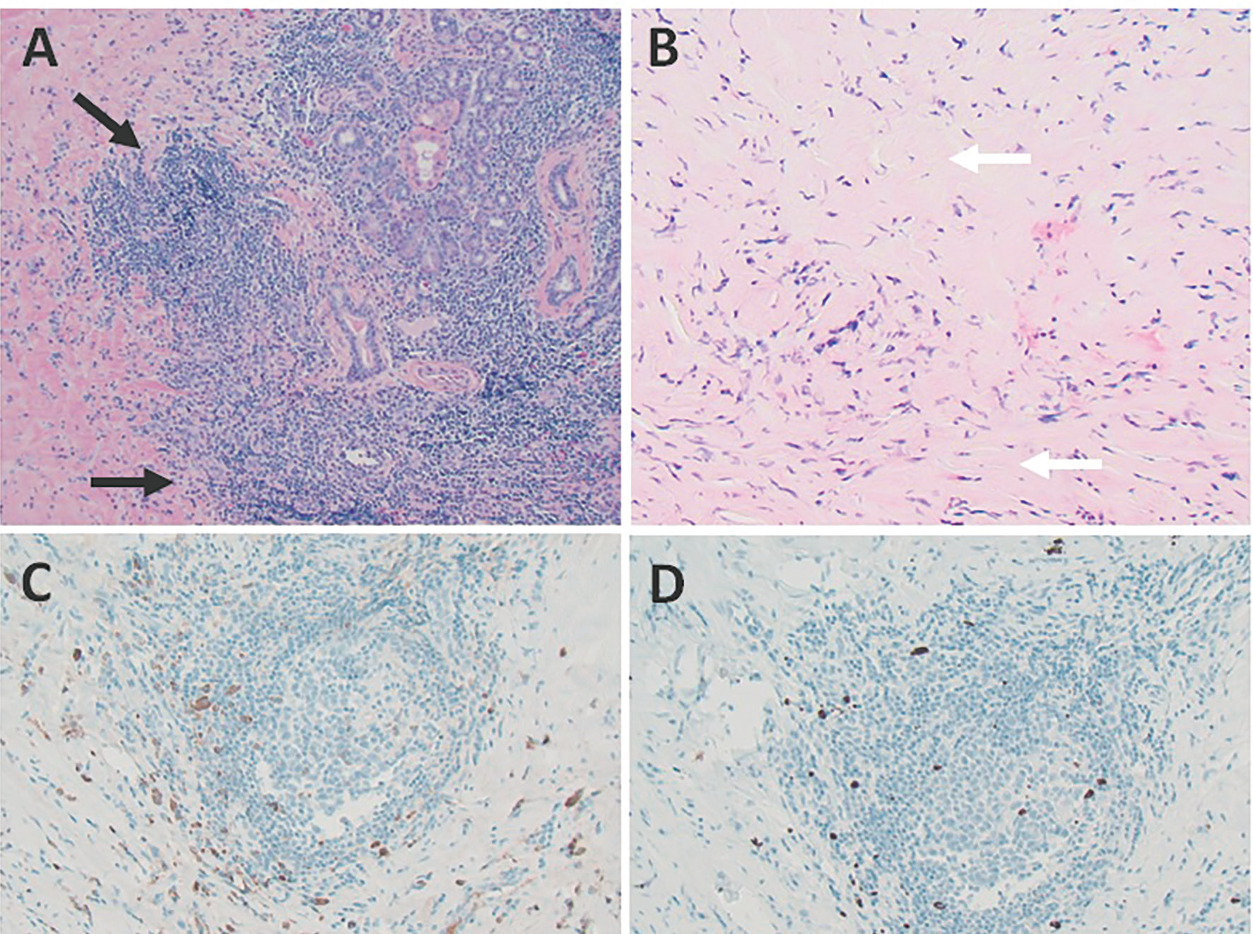
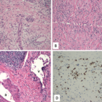
FIGURES 2 A, B, C & D: Right submandibular gland biopsy: (A) H&E staining with marked lymphoplasmacytic infiltrates (black arrows) without evidence of obliterative phlebitis; (B) fibrosis (white arrows) arranged in a swirling pattern known as storiform fibrosis, which is a hallmark feature of IgG4-RD; immunohistochemistry showed focally increased IgG+ (brown cytoplasmic

staining); (C) IgG4+ (brown cytoplasmic staining); and (D) plasma cells with a ratio of ~4:1. The key histologic features of IgG4-RD are dense lymphoplasmacytic infiltrate, storiform-patterned fibrosis, obliterative phlebitis and infiltration of IgG4+ plasma cells with ratio of

[mobile-ad name="Advert 2"]

IgG4/IgG cells >40%, and >10 IgG4+ plasma cells/high-powered field in biopsy tissue sample. (Click to enlarge.)

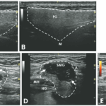
Ultrasonography findings in IgG4-RD and Sjögren’s disease show both similarities and differences. A reticular pattern can be observed in both conditions, but a distinct nodal pattern is more common in the submandibular glands of IgG4-RD and can help distinguish it from Sjögren’s disease.7 This nodal pattern is characterized by hypoechoic, focal, homogenous areas with increased vascularity and protrusion from the surface of the glands. Submandibular glands are predominantly affected in IgG4-RD, while the involvement of parotid glands is less frequent.6 The presence of focal, hypoechoic nodular lesions primarily affecting the submandibular glands while sparing the parotid glands may indicate IgG4-RD.

Page: 1 2 3 4 | Single Page
Share: 

Filed under:ConditionsOther Rheumatic ConditionsSjögren’s Disease Tagged with:case reportglandular swellingIgG4 related diseaseimage case reportSalivary gland ultrasonographySjogren'sUltrasound

Related Articles
    IgG4-Related Kidney Disease: Diagnostics, Manifestations, & More

    IgG4-Related Kidney Disease: Diagnostics, Manifestations & More

    May 17, 2018

    Immunoglobin G4-related disease (IgG4-RD) is a rare fibro-inflammatory disease of unknown etiology that has been recently recognized. It can cause fibro-inflammatory masses in almost every organ of the body and is associated with dense lymphoplasmacytic infiltration of IgG4-postitive plasma cells, storiform fibrosis and elevated levels of serum IgG4.1 IgG4-RD is a systemic disease that may…

    A Spotlight on IgG4-Related Disease

    January 1, 2013

    What rheumatologists need to know about identifying and diagnosing immunoglobulin G4-related disease (IgG4-RD)

    Case Report: Sarcoidosis in Patient with History of IgG4-Related Disease

    September 14, 2021

    Sarcoidosis and IgG4-related disease (IgG4-RD) are both immune-mediated, often multi-organ, diseases of uncertain etiology capable of presenting with diverse clinical manifestations. Many clinical features are common to both conditions, including hypergammaglobulinemia, the ability to form inflammatory masses and involvement of the lymph nodes, lacrimal glands, salivary glands, meninges and lungs. Although imaging modalities, such as…

    Case Report: A Patient with Submandibular Swelling

    March 15, 2021

    We report on a 77-year-old patient who was diagnosed with giant cell arteritis (GCA) on histopathology of the submandibular gland and eventually went on to develop classic signs and symptoms of GCA. This observation highlights neck swelling as a rare initial presentation of GCA. Case Presentation A 77-year-old previously healthy man presented to his primary…

  • About Us
  • Meet the Editors
  • Issue Archives
  • Contribute
  • Advertise
  • Contact Us
  • Copyright © 2025 by John Wiley & Sons, Inc. All rights reserved, including rights for text and data mining and training of artificial technologies or similar technologies. ISSN 1931-3268 (print). ISSN 1931-3209 (online).
  • DEI Statement
  • Privacy Policy
  • Terms of Use
  • Cookie Preferences