Video: Every Case Tells a Story| Webinar: ACR/CHEST ILD Guidelines in Practice

An official publication of the ACR and the ARP serving rheumatologists and rheumatology professionals

  • Conditions
    • Axial Spondyloarthritis
    • Gout and Crystalline Arthritis
    • Myositis
    • Osteoarthritis and Bone Disorders
    • Pain Syndromes
    • Pediatric Conditions
    • Psoriatic Arthritis
    • Rheumatoid Arthritis
    • Sjögren’s Disease
    • Systemic Lupus Erythematosus
    • Systemic Sclerosis
    • Vasculitis
    • Other Rheumatic Conditions
  • FocusRheum
    • ANCA-Associated Vasculitis
    • Axial Spondyloarthritis
    • Gout
    • Psoriatic Arthritis
    • Rheumatoid Arthritis
    • Systemic Lupus Erythematosus
  • Guidance
    • Clinical Criteria/Guidelines
    • Ethics
    • Legal Updates
    • Legislation & Advocacy
    • Meeting Reports
      • ACR Convergence
      • Other ACR meetings
      • EULAR/Other
    • Research Rheum
  • Drug Updates
    • Analgesics
    • Biologics/DMARDs
  • Practice Support
    • Billing/Coding
    • EMRs
    • Facility
    • Insurance
    • QA/QI
    • Technology
    • Workforce
  • Opinion
    • Patient Perspective
    • Profiles
    • Rheuminations
      • Video
    • Speak Out Rheum
  • Career
    • ACR ExamRheum
    • Awards
    • Career Development
  • ACR
    • ACR Home
    • ACR Convergence
    • ACR Guidelines
    • Journals
      • ACR Open Rheumatology
      • Arthritis & Rheumatology
      • Arthritis Care & Research
    • From the College
    • Events/CME
    • President’s Perspective
  • Search

Image Case Report: Refractory, Acute, Cutaneous Lupus

Samantha C. Shapiro, MD  |  Issue: June 2022  |  June 14, 2022

A 25-year-old Mexican American woman with a five-year history of systemic lupus erythematosus (SLE) presents with refractory, acute cutaneous lupus erythematosus (ACLE) and subacute cutaneous lupus erythematosus (SCLE) affecting the scalp, face and hands. Her serologic phenotype is characterized by elevated anti-nuclear, anti-double-stranded deoxyribonucleic acid (dsDNA), anti-ribonucleoprotein (RNP), anti-Smith and anti-SS-A (Ro) antibodies and chronically low serum complement levels.

Her clinical phenotype is characterized by ACLE, SCLE with features of Rowell syndrome (i.e., erythema multiforme-like lesions), arthralgia, oral ulcers, leukopenia and thrombocytopenia. She had no internal organ manifestations of disease.

ad goes here:advert-1
ADVERTISEMENT
SCROLL TO CONTINUE

Her cutaneous disease was refractory to multiple medications. Systemic therapies included 400 mg of hydroxychloroquine by mouth daily, 25 mg of subcutaneous methotrexate weekly and 1,500 mg of mycophenolate mofetil by mouth twice daily. Adjunctive topical corticosteroids and topical calcineurin inhibitors were ineffective. She had required prednisone since diagnosis, with doses ranging from 5 mg to 60 mg by mouth daily. When the prednisone was tapered below 10 mg by mouth daily, cutaneous disease flared.

Clinical Findings

The physical exam was notable for active ACLE and SCLE lesions despite adherence to combination therapy with hydroxychloroquine, mycophenolate mofetil, methotrexate, prednisone 10 mg daily and topicals. A classic malar butterfly rash of the forehead, chin and malar cheeks, with relative sparing of the nasolabial folds, was present (see Figure 1). Significant involvement of the posterior scalp and ears was seen, with residual scarring alopecia (see Figure 2). A maculopapular, erythematous and scaling rash presented on the dorsal and palmar surfaces of bilateral hands (see Figures 3 & 4). These photographs were obtained at her most recent clinic visit.
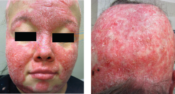
Classic malar butterfly rash of the forehead, chin and malar cheeks. Note the relative sparing of the nasolabial folds.

Figures 1 & 2. Classic malar butterfly rash of the forehead, chin and malar cheeks. Note the relative sparing of the nasolabial folds.

Discussion

Cutaneous involvement in SLE is common and heterogeneous, with rashes ranging from mild to severe and refractory. A predilection for sun-exposed areas exists, and photosensitivity is common. First-line treatment for ACLE includes sun protection, smoking cessation, topical therapies and antimalarial agents (e.g., hydroxychloroquine, chloroquine and quinacrine). For many patients, this approach alone may result in dramatic improvement, with one recent meta-analysis demonstrating significant response in 91% of ACLE cases.1

However, some patients may have refractory cutaneous disease, requiring augmented immunosuppression. Methotrexate and mycophenolate mofetil are both reasonable second-line treatment options. Azathioprine may also be considered, but is generally less efficacious than the aforementioned treatments.2

Our patient tried and failed maximum doses of both methotrexate and myco­phenolate mofetil on a background of hydroxychloroquine, as well as a brief trial of combination methotrexate, mycophenolate mofetil and hydroxychloroquine with careful lab monitoring for toxicity.
Page: 1 2 | Single Page
Share: 

Filed under:ConditionsSystemic Lupus Erythematosus Tagged with:case reportcutaneous lupus erythematosus (CLE)diagnostic imagingimage caseimage case report

Related Articles

    Dermatologist, Rheumatologist Discuss Refractory Cutaneous Lupus Case

    September 5, 2022

    As a dermatologist/internist with a career-long subspecialty interest in the cutaneous manifestations of the rheumatic diseases, I found the case of refractory acute cutaneous lupus by Samantha C. Shapiro, MD, in the June 2022 issue of The Rheumatologist intriguing in several ways, and I felt my perspectives on this case might provide additional educational value…

    nukeaf / shutterstock.com

    The History of Treating Lupus with Hydroxychloroquine

    June 15, 2020

    Given how unexpectedly front and center hydroxychloroquine has been in discussions about the treatment of COVID-19 this year, it makes sense to look at how it became so central to the treatment of a rheumatologic condition. In 1991, an article appeared in The New England Journal of Medicine that would alter the way rheumatologists approached…

    2015 ACR/ARHP Annual Meeting: Skin Issues in Rheumatic Diseases Present Challenges

    March 15, 2016

    SAN FRANCISCO—A 40-year-old woman shows up in the clinic with scarring alopecia, with an area of hyperpigmentation on the rim of her scalp, extending from just behind the temple to behind her ears. An examination with a dermatoscope shows hyperkeratotic follicular plugging. ad goes here:advert-1ADVERTISEMENTSCROLL TO CONTINUEThe case—in this example, the discoid form of cutaneous…

    Dermatology & Immunology: Skin Issues Can Present Challenges

    February 23, 2016

    SAN FRANCISCO—A 40-year-old woman shows up in the clinic with scarring alopecia, with an area of hyperpigmentation on the rim of her scalp, extending from just behind the temple to behind her ears. An examination with a dermatoscope shows hyperkeratotic follicular plugging. ad goes here:advert-1ADVERTISEMENTSCROLL TO CONTINUEThe case—in this example, the discoid form of cutaneous…

  • About Us
  • Meet the Editors
  • Issue Archives
  • Contribute
  • Advertise
  • Contact Us
  • Copyright © 2025 by John Wiley & Sons, Inc. All rights reserved, including rights for text and data mining and training of artificial technologies or similar technologies. ISSN 1931-3268 (print). ISSN 1931-3209 (online).
  • DEI Statement
  • Privacy Policy
  • Terms of Use
  • Cookie Preferences