Video: Every Case Tells a Story| Webinar: ACR/CHEST ILD Guidelines in Practice

An official publication of the ACR and the ARP serving rheumatologists and rheumatology professionals

  • Conditions
    • Axial Spondyloarthritis
    • Gout and Crystalline Arthritis
    • Myositis
    • Osteoarthritis and Bone Disorders
    • Pain Syndromes
    • Pediatric Conditions
    • Psoriatic Arthritis
    • Rheumatoid Arthritis
    • Sjögren’s Disease
    • Systemic Lupus Erythematosus
    • Systemic Sclerosis
    • Vasculitis
    • Other Rheumatic Conditions
  • FocusRheum
    • ANCA-Associated Vasculitis
    • Axial Spondyloarthritis
    • Gout
    • Psoriatic Arthritis
    • Rheumatoid Arthritis
    • Systemic Lupus Erythematosus
  • Guidance
    • Clinical Criteria/Guidelines
    • Ethics
    • Legal Updates
    • Legislation & Advocacy
    • Meeting Reports
      • ACR Convergence
      • Other ACR meetings
      • EULAR/Other
    • Research Rheum
  • Drug Updates
    • Analgesics
    • Biologics/DMARDs
  • Practice Support
    • Billing/Coding
    • EMRs
    • Facility
    • Insurance
    • QA/QI
    • Technology
    • Workforce
  • Opinion
    • Patient Perspective
    • Profiles
    • Rheuminations
      • Video
    • Speak Out Rheum
  • Career
    • ACR ExamRheum
    • Awards
    • Career Development
  • ACR
    • ACR Home
    • ACR Convergence
    • ACR Guidelines
    • Journals
      • ACR Open Rheumatology
      • Arthritis & Rheumatology
      • Arthritis Care & Research
    • From the College
    • Events/CME
    • President’s Perspective
  • Search

Inflammatory Myopathies Difficult to Diagnose, Treat

Jonathan Jones, MD, and Robert Wortmann, MD  |  Issue: December 2013  |  December 1, 2013

The Challenge of Inflammatory Myopathies

ad goes here:advert-1
ADVERTISEMENT
SCROLL TO CONTINUE

Rheumatologists often have varying levels of comfort in assessing and managing muscle disease. For many, these diseases are challenging because of the difficulty in accurately diagnosing myopathies and establishing effective therapeutic regimens. We outline three key issues in diagnosing and managing inflammatory myopathies.

No Gold Standard for Diagnosis

Because there is no gold standard for diagnosing idiopathic inflammatory myopathies, we are forced to rely on criteria. Although more than a dozen sets of criteria have been proposed, the first set, published in 1975 by Bohan and Peter, remains the mainstay.1,2 For the diagnosis of polymyositis, they proposed the combination of proximal muscle weakness, elevated serum levels of enzymes derived from skeletal muscle, a classic triad of electromyographic (EMG) abnormalities, and the presence of inflammation on muscle biopsy. The diagnosis of dermatomyositis could be rendered with the addition of a characteristic skin rash. These parameters seem clear cut until one gets into the details, because each criterion by itself is very nonspecific. For example, fixed proximal muscle weakness may be the hallmark of inflammatory myopathies, but this symptom may be caused by a myriad of other conditions (see Table 1).

ad goes here:advert-2
ADVERTISEMENT
SCROLL TO CONTINUE

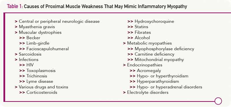
When elevated, the enzyme most considered to be a sign of myositis is creatine phosphokinase (CPK). It is sometimes mistakenly believed that CPK must be elevated at least one time during the course of polymyositis, but this is not always the case. Some patients have normal CPK levels throughout their course. Furthermore, there are several other causes of an elevated CPK, including exercise (anaerobic and aerobic), trauma (blunt or sharp), drugs (statins, colchicine, alcohol, cocaine), and toxins. Carriers for muscular dystrophy or some metabolic myopathies may have an increased serum CPK. Healthy African-American males commonly have CPK levels above the upper limits of normal reported for the population as a whole. Finally, in large clinical trials using statin drugs, over 30% of individuals have elevated CPK levels. Interestingly, the percentage was the same for those taking placebo as it was for those taking study drugs.3 Thus, relying on this test as a way to screen for muscle inflammation can be problematic.

Causes of Proximal Muscle Weakness That May Mimic Inflammatory Myopathy
click for large version
Table 1: Causes of Proximal Muscle Weakness That May Mimic Inflammatory Myopathy

The classic triad of EMG changes described includes: 1) fibrillation at rest, increased insertional activity, and spontaneous and positive sharp waves; 2) bizarre high-frequency repetitive discharges; and 3) polyphasic potentials of short duration and low amplitude.1,2 However, this triad is not diagnostic and is generally present in only 40% of patients with myositis, while 10% of patients with these diseases have normal EMG results.4

Page: 1 2 3 4 5 | Single Page
Share: 

Filed under:ConditionsMyositisOther Rheumatic Conditions Tagged with:dermatomyositisDiagnosisinflammatory myopathymyositispolymyositisrheumatologyTreatment

Related Articles

    New Tools for Myositis Diagnosis, Classification & Management

    April 15, 2019

    CHICAGO—At Hot Topics in Myositis, a session at the 2018 ACR/ARHP Annual Meeting, three experts discussed new classification criteria for idiopathic inflammatory myopathies (IIM) and offered practical primers on overlap myositis conditions and inclusion body myositis (IBM). New Myositis Classification Criteria After a 10-year development process, the new EULAR/ACR Classification Criteria for Adult and Juvenile…

    Fellows’ Forum Case Report: Necrotizing Autoimmune Myopathy

    December 18, 2017

    Necrotizing autoimmune myopathy (NAM) is a relatively recently discovered subgroup of inflammatory myopathies. NAM is characterized by predominant muscle fiber necrosis and regeneration with little or no inflammation.1 One subgroup of NAM is 3-hydroxy-3-methylglutaryl-CoA reductase antibody (HMGCR Ab)-related immune-mediated necrotizing myopathy (IMNM), which occurs (rarely) after statin exposure, with a rough incidence of two per…

    Myositis Mysteries

    January 1, 2008

    Why isn’t my myositis patient getting better?

    Diagnosis: Myopathy

    July 1, 2009

    Presentation and evaluation of metabolic causes

  • About Us
  • Meet the Editors
  • Issue Archives
  • Contribute
  • Advertise
  • Contact Us
  • Copyright © 2025 by John Wiley & Sons, Inc. All rights reserved, including rights for text and data mining and training of artificial technologies or similar technologies. ISSN 1931-3268 (print). ISSN 1931-3209 (online).
  • DEI Statement
  • Privacy Policy
  • Terms of Use
  • Cookie Preferences