Video: Every Case Tells a Story| Webinar: ACR/CHEST ILD Guidelines in Practice

An official publication of the ACR and the ARP serving rheumatologists and rheumatology professionals

  • Conditions
    • Axial Spondyloarthritis
    • Gout and Crystalline Arthritis
    • Myositis
    • Osteoarthritis and Bone Disorders
    • Pain Syndromes
    • Pediatric Conditions
    • Psoriatic Arthritis
    • Rheumatoid Arthritis
    • Sjögren’s Disease
    • Systemic Lupus Erythematosus
    • Systemic Sclerosis
    • Vasculitis
    • Other Rheumatic Conditions
  • FocusRheum
    • ANCA-Associated Vasculitis
    • Axial Spondyloarthritis
    • Gout
    • Psoriatic Arthritis
    • Rheumatoid Arthritis
    • Systemic Lupus Erythematosus
  • Guidance
    • Clinical Criteria/Guidelines
    • Ethics
    • Legal Updates
    • Legislation & Advocacy
    • Meeting Reports
      • ACR Convergence
      • Other ACR meetings
      • EULAR/Other
    • Research Rheum
  • Drug Updates
    • Analgesics
    • Biologics/DMARDs
  • Practice Support
    • Billing/Coding
    • EMRs
    • Facility
    • Insurance
    • QA/QI
    • Technology
    • Workforce
  • Opinion
    • Patient Perspective
    • Profiles
    • Rheuminations
      • Video
    • Speak Out Rheum
  • Career
    • ACR ExamRheum
    • Awards
    • Career Development
  • ACR
    • ACR Home
    • ACR Convergence
    • ACR Guidelines
    • Journals
      • ACR Open Rheumatology
      • Arthritis & Rheumatology
      • Arthritis Care & Research
    • From the College
    • Events/CME
    • President’s Perspective
  • Search

Inflammatory Myopathies Difficult to Diagnose, Treat

Jonathan Jones, MD, and Robert Wortmann, MD  |  Issue: December 2013  |  December 1, 2013

Myositis-Specific Autoantibodies
click for large version
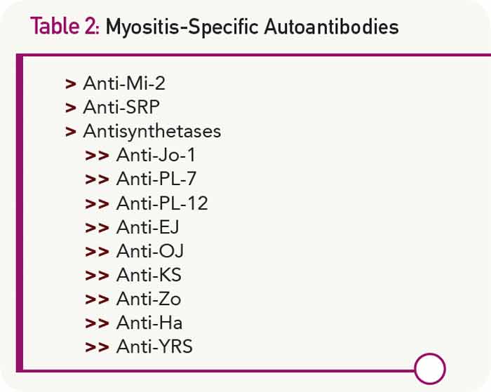
Table 2: Myositis-Specific Autoantibodies

The Differential Diagnosis Is Huge

Another challenge in diagnosing idiopathic inflammatory myopathies involves differentiating them from many other conditions, considered to be myositis mimics, which can also fulfill Bohan and Peter’s criteria (see Table 1). Bohan and Peter recognized the relative nonspecific nature of their criteria, stating, “There are certain findings, herein called exclusions, whose presence should make one very cautious about rendering a diagnosis of polymyositis.”2

ad goes here:advert-1
ADVERTISEMENT
SCROLL TO CONTINUE

The first group of myositis mimics are muscular dystrophies. These are inherited disorders characterized by progressive muscle weakness that advances to muscle wasting. Becker, limb-girdle, and facioscapular dystrophies may present in late adolescence or adulthood with proximal muscle weakness and elevated muscle enzymes. Although the classic histologic picture in these diseases includes muscle-fiber size variation with necrosis and eventually replacement of muscle by fat and fibrous tissue, inflammatory cells may also be observed in early stages. The situation may be further complicated by the observation that some patients with muscular dystrophy note a temporary improvement with corticosteroid therapy.

Selected infections can cause a chronic myopathy. This is particularly true for immunocompromised individuals in whom toxoplasmosis infection can result in the development of an inflammatory myopathy. Some patients infected with HIV develop a slowly progressive proximal myopathy with elevated CPK levels. Muscle biopsy demonstrates endomysial, perimysial, or perivascular mononuclear cell infiltrates and muscle fiber necrosis.

ad goes here:advert-2
ADVERTISEMENT
SCROLL TO CONTINUE

Many drugs can induce a myopathy. Corticosteroids and HMG-CoA reductase inhibitors (statins) are among the most common. Corticosteroid use can result in the development of painless proximal muscle weakness, especially involving the pelvic girdle muscles and neck flexors. In patients being treated for diseases other than myositis, the serum CPK is normal and magnetic resonance imaging of the muscles is unremarkable. EMG may show low-amplitude motor unit potentials, and muscle biopsy classically demonstrates type 2 fiber atrophy. The clinical picture may be confounding when a myositis patient treated with corticosteroids initially improves to a degree and then begins to regress. Is the myositis flaring, or is there a superimposed corticosteroid myopathy? Generally, the latter condition occurs in patients who have been treated with corticosteroids for at least four weeks or, in most cases, for much longer. It is more likely to occur in patients treated with higher doses of corticosteroids, especially the fluorinated preparations (triamcinolone, dexamethasone, or betamethasone). One strategy to diagnose steroid myopathy is to rapidly taper the dose and see if there is clinical improvement or worsening.

Page: 1 2 3 4 5 | Single Page
Share: 

Filed under:ConditionsMyositisOther Rheumatic Conditions Tagged with:dermatomyositisDiagnosisinflammatory myopathymyositispolymyositisrheumatologyTreatment

Related Articles

    New Tools for Myositis Diagnosis, Classification & Management

    April 15, 2019

    CHICAGO—At Hot Topics in Myositis, a session at the 2018 ACR/ARHP Annual Meeting, three experts discussed new classification criteria for idiopathic inflammatory myopathies (IIM) and offered practical primers on overlap myositis conditions and inclusion body myositis (IBM). New Myositis Classification Criteria After a 10-year development process, the new EULAR/ACR Classification Criteria for Adult and Juvenile…

    Fellows’ Forum Case Report: Necrotizing Autoimmune Myopathy

    December 18, 2017

    Necrotizing autoimmune myopathy (NAM) is a relatively recently discovered subgroup of inflammatory myopathies. NAM is characterized by predominant muscle fiber necrosis and regeneration with little or no inflammation.1 One subgroup of NAM is 3-hydroxy-3-methylglutaryl-CoA reductase antibody (HMGCR Ab)-related immune-mediated necrotizing myopathy (IMNM), which occurs (rarely) after statin exposure, with a rough incidence of two per…

    Myositis Mysteries

    January 1, 2008

    Why isn’t my myositis patient getting better?

    Diagnosis: Myopathy

    July 1, 2009

    Presentation and evaluation of metabolic causes

  • About Us
  • Meet the Editors
  • Issue Archives
  • Contribute
  • Advertise
  • Contact Us
  • Copyright © 2025 by John Wiley & Sons, Inc. All rights reserved, including rights for text and data mining and training of artificial technologies or similar technologies. ISSN 1931-3268 (print). ISSN 1931-3209 (online).
  • DEI Statement
  • Privacy Policy
  • Terms of Use
  • Cookie Preferences