Video: Every Case Tells a Story| Webinar: ACR/CHEST ILD Guidelines in Practice

An official publication of the ACR and the ARP serving rheumatologists and rheumatology professionals

  • Conditions
    • Axial Spondyloarthritis
    • Gout and Crystalline Arthritis
    • Myositis
    • Osteoarthritis and Bone Disorders
    • Pain Syndromes
    • Pediatric Conditions
    • Psoriatic Arthritis
    • Rheumatoid Arthritis
    • Sjögren’s Disease
    • Systemic Lupus Erythematosus
    • Systemic Sclerosis
    • Vasculitis
    • Other Rheumatic Conditions
  • FocusRheum
    • ANCA-Associated Vasculitis
    • Axial Spondyloarthritis
    • Gout
    • Psoriatic Arthritis
    • Rheumatoid Arthritis
    • Systemic Lupus Erythematosus
  • Guidance
    • Clinical Criteria/Guidelines
    • Ethics
    • Legal Updates
    • Legislation & Advocacy
    • Meeting Reports
      • ACR Convergence
      • Other ACR meetings
      • EULAR/Other
    • Research Rheum
  • Drug Updates
    • Analgesics
    • Biologics/DMARDs
  • Practice Support
    • Billing/Coding
    • EMRs
    • Facility
    • Insurance
    • QA/QI
    • Technology
    • Workforce
  • Opinion
    • Patient Perspective
    • Profiles
    • Rheuminations
      • Video
    • Speak Out Rheum
  • Career
    • ACR ExamRheum
    • Awards
    • Career Development
  • ACR
    • ACR Home
    • ACR Convergence
    • ACR Guidelines
    • Journals
      • ACR Open Rheumatology
      • Arthritis & Rheumatology
      • Arthritis Care & Research
    • From the College
    • Events/CME
    • President’s Perspective
  • Search

Malignant Atrophic Papulosis Is Challenging to Diagnose, Treat

Aixa Toledo-Garcia, MD, Jessica Farrell, PharmD, & Lee Shapiro, MD  |  Issue: July 2014  |  July 1, 2014

Figure 2: An exploratory laparotomy revealed multiple ulcerated lesions throughout the small bowel.

A few months later, the patient developed a small bowel obstruction as a result of adhesions. His hospitalization was complicated by the development of pleural effusion requiring thoracentesis. He also experienced a 30 lb. weight loss and severe deconditioning, requiring total parenteral nutrition for bowel rest. Additionally, he developed a new supraventricular tachycardia.

ad goes here:advert-1
ADVERTISEMENT
SCROLL TO CONTINUE

Once again the search for answers started.

ad goes here:advert-2
ADVERTISEMENT
SCROLL TO CONTINUE
Figure 3: Cystoscopy demonstrated multiple lesions suggestive of MAP.

Figure 4: Left: Extensive avascular lesions with telangectatic borders on a second patient’s digits and hands before treatment with treprostinil. Right: Her hands after treatment with treprostinil.

Figure 5: Left: MRI imaging shows the T1 postcontrast image demonstrating the cord lesion in the<br /><br /><br />
left frontal lobe in December 2010. Right: On repeat imaging in July 2011, the enhancement is mostly resolved.
Figure 5: Left: MRI imaging shows the T1 postcontrast image demonstrating the cord lesion in the left frontal lobe in December 2010. Right: On repeat imaging in July 2011, the enhancement is mostly resolved.

Lesson Learned from a 2nd Patient

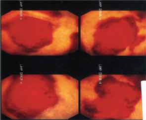
With the unique nature of MAP skin lesions, we were able to identify similar lesions on another patient previously diagnosed with an overlap syndrome with features of systemic lupus erythematous and systemic sclerosis. The skin lesions were biopsied and were reported as MAP-like lesions. Subsequently, the patient was started on subcutaneous treprostinil for treatment of her severe pulmonary hypertension. Interestingly, within three months of initiating therapy with treprostinil, her MAP-like skin lesions showed marked involution (see Figure 4).

Based on these findings, in December 2010, we were able to obtain treprostinil on a compassionate-use basis for our 17-year-old male patient. Slowly, he improved and has been maintained on a regimen of treprostinil, eculizumab and aspirin.

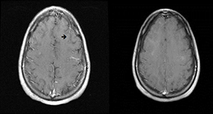
Repeat MRI of the brain showed complete resolution of brain lesions (see Figure 5). He continues to develop some skin lesions, but they involute more quickly and remain smaller in size compared with presentation prior to treatment.

A 3rd Patient

Our 17-year-old patient’s sister was found to have biopsy-proven MAP skin lesions. She underwent exploratory laparoscopy to evaluate complaints of abdominal pain, and no serosal lesions were seen. The pain spontaneously resolved. It appears that she has cutaneous disease only.

What Is Malignant Atrophic Papulosis?

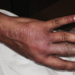
MAP is an uncommon thrombo-occlusive vascu­lopathy with characteristic skin lesions.1 Similar lesions can affect internal organs, such as the GI tract, central nervous system, pericardium and bladder.2 Thrombotic complications with characteristic infarcted zones can be seen on histopathology.3,4 Cutaneous-only involvement has been described. Conversely, previously reported systemic disease has been fatal.

Page: 1 2 3 4 5 | Single Page
Share: 

Filed under:Biologics/DMARDsConditionsDrug UpdatesSystemic Lupus ErythematosusSystemic SclerosisVasculitis Tagged with:Biologicsdruginfliximabintravenous immune globulinpatient carerheumatologistSclerodermasystemic lupus erythematousVasculitis

Related Articles

    Looking for Links Between Rheumatoid Arthritis & Gut Bacteria, Mutations

    June 21, 2018

    Patients with a specific genetic predisposition for rheumatoid arthritis (RA) may want to take precautions when consuming certain types of meat, dairy and produce grown with cow manure, according to a new study out of the University of Central Florida (UCF) in Orlando. The study, which assessed 100 RA patients, published in the journal Frontiers…

    The Science of Chronic Itch

    December 1, 2014

    Inside the pathophysiology, clinical presentations of chronic pruritus

    Rheumatology Case Report: Bullous Lesions in Patient with Lupus

    April 20, 2017

    Systemic lupus erythematosus (SLE) is a heterogeneous disease associated with multiple acute or chronic cutaneous manifestations, including the relatively rare category of bullous lupus. The development of vesiculo-bullous lesions may be associated with a high morbidity, hence they warrant an urgent investigation, including a skin biopsy to identify the diagnosis and initiate prompt treatment. With…

    Dermatologist, Rheumatologist Discuss Refractory Cutaneous Lupus Case

    September 5, 2022

    As a dermatologist/internist with a career-long subspecialty interest in the cutaneous manifestations of the rheumatic diseases, I found the case of refractory acute cutaneous lupus by Samantha C. Shapiro, MD, in the June 2022 issue of The Rheumatologist intriguing in several ways, and I felt my perspectives on this case might provide additional educational value…

  • About Us
  • Meet the Editors
  • Issue Archives
  • Contribute
  • Advertise
  • Contact Us
  • Copyright © 2025 by John Wiley & Sons, Inc. All rights reserved, including rights for text and data mining and training of artificial technologies or similar technologies. ISSN 1931-3268 (print). ISSN 1931-3209 (online).
  • DEI Statement
  • Privacy Policy
  • Terms of Use
  • Cookie Preferences