The Rheumatologist
COVID-19 News
  • Connect with us:
  • Facebook
  • Twitter
  • LinkedIn
  • YouTube
  • Feed
  • Home
  • Conditions
    • Rheumatoid Arthritis
    • SLE (Lupus)
    • Crystal Arthritis
      • Gout Resource Center
    • Spondyloarthritis
    • Osteoarthritis
    • Soft Tissue Pain
    • Scleroderma
    • Vasculitis
    • Systemic Inflammatory Syndromes
    • Guidelines
  • Resource Centers
    • Axial Spondyloarthritis Resource Center
    • Gout Resource Center
    • Psoriatic Arthritis Resource Center
    • Rheumatoid Arthritis Resource Center
    • Systemic Lupus Erythematosus Resource Center
  • Drug Updates
    • Biologics & Biosimilars
    • DMARDs & Immunosuppressives
    • Topical Drugs
    • Analgesics
    • Safety
    • Pharma Co. News
  • Professional Topics
    • Ethics
    • Legal
    • Legislation & Advocacy
    • Career Development
      • Certification
      • Education & Training
    • Awards
    • Profiles
    • President’s Perspective
    • Rheuminations
    • Interprofessional Perspective
  • Practice Management
    • Billing/Coding
    • Quality Assurance/Improvement
    • Workforce
    • Facility
    • Patient Perspective
    • Electronic Health Records
    • Apps
    • Information Technology
    • From the College
    • Multimedia
      • Audio
      • Video
  • Resources
    • Issue Archives
    • ACR Convergence
      • Systemic Lupus Erythematosus Resource Center
      • Rheumatoid Arthritis Resource Center
      • Gout Resource Center
      • Abstracts
      • Meeting Reports
      • ACR Convergence Home
    • American College of Rheumatology
    • ACR ExamRheum
    • Research Reviews
    • ACR Journals
      • Arthritis & Rheumatology
      • Arthritis Care & Research
      • ACR Open Rheumatology
    • Rheumatology Image Library
    • Treatment Guidelines
    • Rheumatology Research Foundation
    • Events
  • About Us
    • Mission/Vision
    • Meet the Authors
    • Meet the Editors
    • Contribute to The Rheumatologist
    • Subscription
    • Contact
  • Advertise
  • Search
You are here: Home / Articles / New Evidence MRI Can Be Used to Monitor Neuromuscular Disease Progression

New Evidence MRI Can Be Used to Monitor Neuromuscular Disease Progression

December 14, 2015 • By Lara C. Pullen, PhD

  • Tweet
  • Email
Print-Friendly Version / Save PDF

James Steidl_Shutterstock_MRI500x270
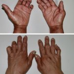
Neuromuscular disorders, such as Charcot-Marie-Tooth type 1A, present many challenges to researchers, one of which is the lack of responsive outcome measures for clinical trials. Without an accurate and reliable means of monitoring disease progression, it becomes difficult for researchers to conduct successful experimental trials of new therapies. In the search for an objective measure, one clue has surfaced: Many of the neuromuscular disorders are characterized by chronic intramuscular fat accumulation that appears to contribute to the clinical presentation.

You Might Also Like
  • No Clear Connection Between Inflammation & Disease Progression in AS
  • Diagnosis Trajectories Aid in Predicting Disease Progression
  • Treating Early-Stage Spondyloarthritis May Prevent Disease Progression, Say Researchers at the ACR/ARHP Winter Rheumatology Symposium
Also By This Author
  • Patients May Be Right: Tomatoes May Trigger Gout Flares

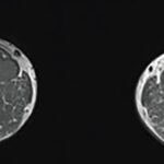
Although different diseases have different distributions and degrees of muscle abnormalities, the presence of the abnormalities and their correlation with symptoms are consistent across multiple neuromuscular disorders. Moreover, several studies have suggested that magnetic resonance imaging (MRI) may be a sensitive and non-invasive method of measuring skeletal muscle disease progression in various neuromuscular diseases. Specifically, investigators have used MRI to evaluate patients with Duchenne muscular dystrophy and have demonstrated that steroid use is associated with a reduction in transverse relaxation time constant (T2) in some patients or a variable effect on T2 in others.

ad goes here:advert-1
ADVERTISEMENT
SCROLL TO CONTINUE

In the case of inclusion body myositis associated with Charcot-Marie-Tooth type 1A, patients first present with minor symptoms. These symptoms progress over approximately 10 years to significant disability. Investigators asked whether it was possible to use MRI to track and measure these changes in muscle. Jasper M. Morrow, FRACP, at the UCL Institute of Neurology in London, and colleagues published the results of their prospective observational cohort study online on Nov. 5 in The Lancet Neurology.1 They evaluated muscle fat fraction using T2 and magnetic transfer ratio (MTR), and compared those measurements with relevant clinical functional tests. The investigators focused their efforts on the thigh and calf muscles of patients with Charcot-Marie-Tooth disease. Specifically, they analyzed data from single slices of thigh and calf blocks, using a small region of interest (ROI) for T2 and MTR sequences.

The researchers reported that MRI is a sensitive measure of disease progression in patients with Charcot-Marie-Tooth disease 1A and inclusion body myositis. Moreover, they found a strong clinical MRI correlation at both the overall participant level, as well as at the level of individual muscles. They also noted that, because the T2 of fat greatly exceeds the T2 of muscle water, T2 is affected by both fat fraction, as well as changes in tissue water distribution. Thus, MRI can be used to detect the muscle water changes that precede marked intramuscular fat accumulation.

ad goes here:advert-2
ADVERTISEMENT
SCROLL TO CONTINUE

Pages: 1 2 | Single Page

Filed Under: Conditions, Scleroderma Tagged With: connective tissue disorder

You Might Also Like:
  • No Clear Connection Between Inflammation & Disease Progression in AS
  • Diagnosis Trajectories Aid in Predicting Disease Progression
  • Treating Early-Stage Spondyloarthritis May Prevent Disease Progression, Say Researchers at the ACR/ARHP Winter Rheumatology Symposium
  • Whole-Body MRI, Ultrasound Imaging May Aid in Early Rheumatic Disease Diagnosis, Treatment

Meeting Abstracts

Browse and search abstracts from the ACR Convergence and ACR/ARP Annual Meetings going back to 2012.

Visit the Abstracts site »

American College of Rheumatology

Visit the official website for the American College of Rheumatology.

Visit the ACR »

Simple Tasks

Learn more about the ACR’s public awareness campaign and how you can get involved. Help increase visibility of rheumatic diseases and decrease the number of people left untreated.

Visit the Simple Tasks site »

The Rheumatologist newsmagazine reports on issues and trends in the management and treatment of rheumatic diseases. The Rheumatologist reaches 11,500 rheumatologists, internists, orthopedic surgeons, nurse practitioners, physician assistants, nurses, and other healthcare professionals who practice, research, or teach in the field of rheumatology.

About Us / Contact Us / Advertise / Privacy Policy / Terms of Use

  • Connect with us:
  • Facebook
  • Twitter
  • LinkedIn
  • YouTube
  • Feed

Copyright © 2006–2022 American College of Rheumatology. All rights reserved.

ISSN 1931-3268 (print)
ISSN 1931-3209 (online)