Video: Every Case Tells a Story| Webinar: ACR/CHEST ILD Guidelines in Practice

An official publication of the ACR and the ARP serving rheumatologists and rheumatology professionals

  • Conditions
    • Axial Spondyloarthritis
    • Gout and Crystalline Arthritis
    • Myositis
    • Osteoarthritis and Bone Disorders
    • Pain Syndromes
    • Pediatric Conditions
    • Psoriatic Arthritis
    • Rheumatoid Arthritis
    • Sjögren’s Disease
    • Systemic Lupus Erythematosus
    • Systemic Sclerosis
    • Vasculitis
    • Other Rheumatic Conditions
  • FocusRheum
    • ANCA-Associated Vasculitis
    • Axial Spondyloarthritis
    • Gout
    • Psoriatic Arthritis
    • Rheumatoid Arthritis
    • Systemic Lupus Erythematosus
  • Guidance
    • Clinical Criteria/Guidelines
    • Ethics
    • Legal Updates
    • Legislation & Advocacy
    • Meeting Reports
      • ACR Convergence
      • Other ACR meetings
      • EULAR/Other
    • Research Rheum
  • Drug Updates
    • Analgesics
    • Biologics/DMARDs
  • Practice Support
    • Billing/Coding
    • EMRs
    • Facility
    • Insurance
    • QA/QI
    • Technology
    • Workforce
  • Opinion
    • Patient Perspective
    • Profiles
    • Rheuminations
      • Video
    • Speak Out Rheum
  • Career
    • ACR ExamRheum
    • Awards
    • Career Development
  • ACR
    • ACR Home
    • ACR Convergence
    • ACR Guidelines
    • Journals
      • ACR Open Rheumatology
      • Arthritis & Rheumatology
      • Arthritis Care & Research
    • From the College
    • Events/CME
    • President’s Perspective
  • Search

Rheumatology Case Report: Bullous Lesions in Patient with Lupus

Mitali Sen, MD, Corrado Minimo, MD, & Ruchika Patel, MD  |  Issue: April 2017  |  April 20, 2017

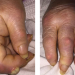
Bullous lupus is relatively rare—described in 1–8% of patients.4,5 Bullous lupus is caused by the development of antibodies against type VII collagen in the basement membrane and manifests with tense, subepidermal vesiculo-bullous eruptions on an erythematous base, more commonly seen on the trunk, face and upper extremities.5 Immunofluorescence reveals continuous or granular pattern of deposition along the dermo-epidermal junction extending to the dermis.

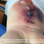
A 2015 clinic-pathological study described blistering lesions in 22 patients with SLE and, based on their histological description, divided them into three groups.6 One group included five patients with extensive epidermal necrosis appearing as TEN-like lesions. A second group of nine patients had classic cutaneous findings, with interface dermatitis with vacuolization of the basal keratinocytes and lymphocytic infiltrate. The third group of eight patients had neutrophilic bullous lesions.

Figure 6: (H&E 60x) Interface vacuolar dermatitis with diffuse acantholysis; note the superficial layer has exfoliated.

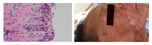
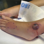
(left) Figure 6: (H&E 60x) Interface vacuolar dermatitis with diffuse acantholysis; note the superficial layer has exfoliated.
(right) Figure 7: Healing lesions after treatment with antibiotics and intravenous immunoglobulins.

SJS and TEN also present as bullous lesions and are dermatological emergencies that usually occur with exposure to new drugs (including disease-modifying anti-rheumatic drugs [DMARDs], such as TNF-alpha inhibitors). However, these conditions usually involve larger body surface area and affect the mucus membranes. Our patient did not have a history of an exposure to a new drug or any other identifiable environmental triggers. She also did not experience any mucosal involvement, making SJS or TENs a less likely possibility.

Studies have described increased morbidity and mortality secondary to infections in SLE patients on immunosuppressive therapy, with a high likelihood of staphylococcal organism involvement due to functional neutrophil dysfunction.7-9 Staphylococcal scalded skin syndrome is caused by an exotoxin released by the bacteria. It attacks the desmosomes, causing detachment of the epidermal layer. It manifests as painful flaccid blisters, positive for Nikolsky’s sign, with widespread desquamation. This disease is more common in children and infants; however, it is associated with mortality rates in adults of approximately 60%, depending on comorbid conditions, degree of immune suppression and delay of treatment initiation.10,11

ad goes here:advert-2
ADVERTISEMENT
SCROLL TO CONTINUE

Staphylococcal scalded skin syndrome causing bullous lesions in a patient with SLE is, however, rare. In our case, we believe the source of infection was possibly the super-infection of the preexisting skin lesions and the nares being the site of colonization for MRSA.

Our review of literature revealed a case of Staphylococcal scalded skin syndrome described in a patient with lupus nephritis, attributed to reduced toxin excretion by the kidneys, as observed in animal models; however, our patient had normal renal function.12 There has also been infrequent case report description of systemic manifestations of toxic shock syndrome without cutaneous involvement in postsurgical or postpartum lupus patients.13,14

Page: 1 2 3 4 5 | Single Page
Share: 

Filed under:ConditionsSystemic Lupus Erythematosus Tagged with:bullous lesioncase reportClinicalcutaneousDiagnosisLupuspatient carerheumatologistrheumatologyscalded skin syndromeSLEstaphylococcalSystemic lupus erythematosusTreatment

Related Articles

    Fellows’ Forum Case Report: Tophaceous Bullae

    August 13, 2017

    As a manifestation of hyperuricemia, inflammatory bullous lesions have rarely been described in the past century. A more classic presentation of hyperuricemia is acute inflammatory gouty arthritis, characterized by the deposition of monosodium urate crystals. Other complications of chronic untreated hyperuricemia may include polyarticular arthritis, tophus formation and possible chronic destructive lesions of the bone,…

    Case Report: A Bullous Eruption

    June 14, 2022

    Eosinophilic granulomatosis with polyangiitis (EGPA) is an anti-neutrophil cytoplasmic antibody-associated vasculitis typically characterized by asthma, peripheral eosinophilia and medium- to small-vessel necrotizing vasculitis. Cutaneous manifes­tations in EGPA are diverse. Palpable purpura is the most common presentation, but urticaria, erythematous macules and papules, livedo reticularis, digital necrosis and cutaneous nodules have also been described.1 Non-hemorrhagic bullae…

    Dermatologist, Rheumatologist Discuss Refractory Cutaneous Lupus Case

    September 5, 2022

    As a dermatologist/internist with a career-long subspecialty interest in the cutaneous manifestations of the rheumatic diseases, I found the case of refractory acute cutaneous lupus by Samantha C. Shapiro, MD, in the June 2022 issue of The Rheumatologist intriguing in several ways, and I felt my perspectives on this case might provide additional educational value…

    Case Report: Sweet Syndrome as an Initial Presentation of Crohn’s Disease

    July 13, 2022

    Acute febrile neutrophilic dermatosis, or Sweet syndrome, is an inflammatory disease that classically presents with fever, leukocytosis and tender, erythematous plaques characterized by neutrophilic infiltrates on biopsy. Sweet syndrome has been reported in association with several autoimmune diseases, including inflammatory bowel disease, systemic lupus erythematous, rheumatoid arthritis and sarcoidosis.1 Here, we discuss a case of…

  • About Us
  • Meet the Editors
  • Issue Archives
  • Contribute
  • Advertise
  • Contact Us
  • Copyright © 2025 by John Wiley & Sons, Inc. All rights reserved, including rights for text and data mining and training of artificial technologies or similar technologies. ISSN 1931-3268 (print). ISSN 1931-3209 (online).
  • DEI Statement
  • Privacy Policy
  • Terms of Use
  • Cookie Preferences