Video: Every Case Tells a Story| Webinar: ACR/CHEST ILD Guidelines in Practice

An official publication of the ACR and the ARP serving rheumatologists and rheumatology professionals

  • Conditions
    • Axial Spondyloarthritis
    • Gout and Crystalline Arthritis
    • Myositis
    • Osteoarthritis and Bone Disorders
    • Pain Syndromes
    • Pediatric Conditions
    • Psoriatic Arthritis
    • Rheumatoid Arthritis
    • Sjögren’s Disease
    • Systemic Lupus Erythematosus
    • Systemic Sclerosis
    • Vasculitis
    • Other Rheumatic Conditions
  • FocusRheum
    • ANCA-Associated Vasculitis
    • Axial Spondyloarthritis
    • Gout
    • Psoriatic Arthritis
    • Rheumatoid Arthritis
    • Systemic Lupus Erythematosus
  • Guidance
    • Clinical Criteria/Guidelines
    • Ethics
    • Legal Updates
    • Legislation & Advocacy
    • Meeting Reports
      • ACR Convergence
      • Other ACR meetings
      • EULAR/Other
    • Research Rheum
  • Drug Updates
    • Analgesics
    • Biologics/DMARDs
  • Practice Support
    • Billing/Coding
    • EMRs
    • Facility
    • Insurance
    • QA/QI
    • Technology
    • Workforce
  • Opinion
    • Patient Perspective
    • Profiles
    • Rheuminations
      • Video
    • Speak Out Rheum
  • Career
    • ACR ExamRheum
    • Awards
    • Career Development
  • ACR
    • ACR Home
    • ACR Convergence
    • ACR Guidelines
    • Journals
      • ACR Open Rheumatology
      • Arthritis & Rheumatology
      • Arthritis Care & Research
    • From the College
    • Events/CME
    • President’s Perspective
  • Search

Rheumatology Case Report: Bullous Lesions in Patient with Lupus

Mitali Sen, MD, Corrado Minimo, MD, & Ruchika Patel, MD  |  Issue: April 2017  |  April 20, 2017

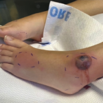
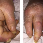
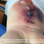
Management of Staphylococcal scalded syndrome includes appropriate antibacterial coverage and intravenous hydration. Given the syndrome’s rarity and severity, this case emphasizes the importance of considering Staphylococcal scalded syndrome in the differential diagnosis of bullous lesions in a patient with SLE.


Mitali Sen, MD, is a second-year internal medicine resident at Albert Einstein Medical Center in Philadelphia.

ad goes here:advert-1
ADVERTISEMENT
SCROLL TO CONTINUE

Corrado Minimo, MD, is the attending physician in the Department of Pathology at Albert Einstein Medical Center in Philadelphia.

Ruchika Patel, MD, is the attending physician in the Department of Rheumatology at Albert Einstein Medical Center in Philadelphia.

ad goes here:advert-2
ADVERTISEMENT
SCROLL TO CONTINUE

References

  1. Crowson A, Magro C. The cutaneous pathology of lupus erythrematosus: A review. J Cutan Pathol. 2001 Jan;28(1):1–23.
  2. Rothfield N, Sontheimer R, Bernstein M, et al. Lupus erythrematosus: Systemic and cutaneous manifestations. Clin Dermatol. 2006 Sep–Oct;24(5):348–362.
  3. Yell JA, Mbuagbaw J, Burge SM. Cutaneous manifestations of systemic lupus erythrematosus. Br J Dermatol. 1996 Sep;135(3):355–362.
  4. Duan L, Chen L, Zhong S, et al. Treatment of bullous systemic lupus erythematosus. J Immunol Res. 2015;2015:167064.
  5. Contestable J, Edgehard K, Meyerle J, et al. Bullous systemic lupus erythrematosus: A review and update to diagnosis and treatment. Am J Clin Dermatol. 2014 Dec;15(6):517–524. doi:10.1007/s40257-014-0098-0.
  6. Merklen-Djafri C, Bessis D, Frances C, et al. Blisters and loss of epidermis in patients with lupus erythematosus: A clinico-pathological study of 22 patients. Medicine (Baltimore). 2015 Nov;94(46):e2102.
  7. Gladman DD, Hussain F, Ibañez D, et al. The nature and outcome of infection in systemic lupus erythematosus. Lupus. 2002;11(4):234–239.
  8. Gresham HD, Ray CJ, O’Sullivan FX. Defective neutrophil function in the autoimmune mouse strain MRL/lpr. Potential role of transforming growth factor-beta. J Immunol. 1991 Jun 1;146(11):3911–3921.
  9. Caver TE, O’Sullivan FX, Gold LI, et al. Intracellular demonstration of active TGFbeta1 in B cells and plasma cells of autoimmune mice. IgG-bound TGFbeta1 suppresses neutrophil function and host defense against Staphylococcus aureus infection. J Clin Invest. 1996 Dec 1;98(11):2496–2506.
  10. Patel GK, Finlay AY. Staphylococcal scalded skin syndrome: Diagnosis and management. Am J Clin Dermatol. 2003;4(3):165–175.
  11. Patel GK. Treatment of staphylococcal scalded skin syndrome. Expert Rev Anti Infect Ther. 2004 Aug;2(4):575–587.
  12. Rydzewska-Rosolowska A, Brzosko S, Borawski J, et al. Staphylococcal scalded skin syndrome in the course of lupus nephritis. Nephrology (Carlton). 2008 Jun;13(3):265–266.
  13. Huseyin TS, Maynard JP, Leach RD. Toxic shock syndrome in a patient with breast cancer and systemic lupus erythematosus. Eur J Surg Oncol. 2001 Apr;27(3):330–331.
  14. Kadoya A, Iikuni Y, Hosaka S, et al. A SLE case with toxic shock syndrome after delivery. Kansenshogaku Zasshi. 1996 Dec;70(12):1279–1783.

Acknowledgments

The authors acknowledge the assistance of the following in the preparation of this article: Sandra Schwarcz, DO, and Jose Nitram-Aliling, MD, fellows in the Department of Rheumatology at Albert Einstein Medical Center, Philadelphia. The pictures were produced with the patient’s permission, and they thank her for allowing the same.
Page: 1 2 3 4 5 | Single Page
Share: 

Filed under:ConditionsSystemic Lupus Erythematosus Tagged with:bullous lesioncase reportClinicalcutaneousDiagnosisLupuspatient carerheumatologistrheumatologyscalded skin syndromeSLEstaphylococcalSystemic lupus erythematosusTreatment

Related Articles

    Fellows’ Forum Case Report: Tophaceous Bullae

    August 13, 2017

    As a manifestation of hyperuricemia, inflammatory bullous lesions have rarely been described in the past century. A more classic presentation of hyperuricemia is acute inflammatory gouty arthritis, characterized by the deposition of monosodium urate crystals. Other complications of chronic untreated hyperuricemia may include polyarticular arthritis, tophus formation and possible chronic destructive lesions of the bone,…

    Case Report: A Bullous Eruption

    June 14, 2022

    Eosinophilic granulomatosis with polyangiitis (EGPA) is an anti-neutrophil cytoplasmic antibody-associated vasculitis typically characterized by asthma, peripheral eosinophilia and medium- to small-vessel necrotizing vasculitis. Cutaneous manifes­tations in EGPA are diverse. Palpable purpura is the most common presentation, but urticaria, erythematous macules and papules, livedo reticularis, digital necrosis and cutaneous nodules have also been described.1 Non-hemorrhagic bullae…

    Dermatologist, Rheumatologist Discuss Refractory Cutaneous Lupus Case

    September 5, 2022

    As a dermatologist/internist with a career-long subspecialty interest in the cutaneous manifestations of the rheumatic diseases, I found the case of refractory acute cutaneous lupus by Samantha C. Shapiro, MD, in the June 2022 issue of The Rheumatologist intriguing in several ways, and I felt my perspectives on this case might provide additional educational value…

    Case Report: Sweet Syndrome as an Initial Presentation of Crohn’s Disease

    July 13, 2022

    Acute febrile neutrophilic dermatosis, or Sweet syndrome, is an inflammatory disease that classically presents with fever, leukocytosis and tender, erythematous plaques characterized by neutrophilic infiltrates on biopsy. Sweet syndrome has been reported in association with several autoimmune diseases, including inflammatory bowel disease, systemic lupus erythematous, rheumatoid arthritis and sarcoidosis.1 Here, we discuss a case of…

  • About Us
  • Meet the Editors
  • Issue Archives
  • Contribute
  • Advertise
  • Contact Us
  • Copyright © 2025 by John Wiley & Sons, Inc. All rights reserved, including rights for text and data mining and training of artificial technologies or similar technologies. ISSN 1931-3268 (print). ISSN 1931-3209 (online).
  • DEI Statement
  • Privacy Policy
  • Terms of Use
  • Cookie Preferences