Video: Every Case Tells a Story| Webinar: ACR/CHEST ILD Guidelines in Practice

An official publication of the ACR and the ARP serving rheumatologists and rheumatology professionals

  • Conditions
    • Axial Spondyloarthritis
    • Gout and Crystalline Arthritis
    • Myositis
    • Osteoarthritis and Bone Disorders
    • Pain Syndromes
    • Pediatric Conditions
    • Psoriatic Arthritis
    • Rheumatoid Arthritis
    • Sjögren’s Disease
    • Systemic Lupus Erythematosus
    • Systemic Sclerosis
    • Vasculitis
    • Other Rheumatic Conditions
  • FocusRheum
    • ANCA-Associated Vasculitis
    • Axial Spondyloarthritis
    • Gout
    • Psoriatic Arthritis
    • Rheumatoid Arthritis
    • Systemic Lupus Erythematosus
  • Guidance
    • Clinical Criteria/Guidelines
    • Ethics
    • Legal Updates
    • Legislation & Advocacy
    • Meeting Reports
      • ACR Convergence
      • Other ACR meetings
      • EULAR/Other
    • Research Rheum
  • Drug Updates
    • Analgesics
    • Biologics/DMARDs
  • Practice Support
    • Billing/Coding
    • EMRs
    • Facility
    • Insurance
    • QA/QI
    • Technology
    • Workforce
  • Opinion
    • Patient Perspective
    • Profiles
    • Rheuminations
      • Video
    • Speak Out Rheum
  • Career
    • ACR ExamRheum
    • Awards
    • Career Development
  • ACR
    • ACR Home
    • ACR Convergence
    • ACR Guidelines
    • Journals
      • ACR Open Rheumatology
      • Arthritis & Rheumatology
      • Arthritis Care & Research
    • From the College
    • Events/CME
    • President’s Perspective
  • Search

Rheumatology Case Report: Chondromyxoid Fibroma of the Sternum

Rajaie Namas, MD, Reshma Khan, MD, & Bernard Rubin, DO  |  Issue: February 2016  |  February 16, 2016

A 47-year-old Caucasian woman presented to the rheumatology clinic with a one-year history of pain and swelling involving the wrists, hands, ankles and feet that progressively worsened. Her symptoms included generalized fatigue, morning stiffness in the hands and lower back lasting more than an hour, Raynaud’s phenomena (triphasic), photosensitivity, tightening of the skin of the hands and feet, heartburn, chest wall tenderness, exertional dyspnea, sicca symptoms and an unintentional weight loss of 30 lbs. over the prior six months.

She had previously been evaluated by another rheumatologist for a four-year history of lower back pain associated with stiffness, which was relieved by non-steroidal anti-inflammatory medications (NSAIDs) and activity. Imaging of the sacroiliac joints revealed bilateral sacroiliitis, and she was diagnosed with ankylosing spondylitis.

ad goes here:advert-1
ADVERTISEMENT
SCROLL TO CONTINUE

One year before, she was also given the diagnosis of scleroderma, due to symmetrical skin thickening distal to the MCPs, synovitis of her MCP and PIP joints, the presence of Raynaud’s phenomenon and a history of esophageal reflux symptoms. She was treated with prednisone 20 mg daily and hydroxychloroquine 200 mg twice daily with minimal improvement in her symptoms.

Medical History

Her medical history was remarkable for hypertension, hypothyroidism and right carpal tunnel release. She was up to date on risk- and age-appropriate malignancy screenings, including mammogram, pap smears and vaccinations.

ad goes here:advert-2
ADVERTISEMENT
SCROLL TO CONTINUE

Social & Family History

The patient was born and raised in Michigan and is happily married with two healthy children. She is employed and is a nonsmoker. She denies chronic alcoholism or illicit drug use. There is no family history of an autoimmune disease.

Physical Examination

At the time of presentation, she was in no acute distress, with a blood pressure of 156/97 mmHg, pulse of 82 beats/min, oxygen saturation of 97% on room air, afebrile and a BMI of 22.9 kg/m2.

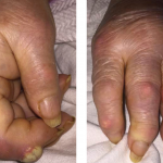
Her physical exam was remarkable for synovitis involving the metacarpophalangeal joints bilaterally, sclerodactyly distal to the MCPs bilaterally, dusky bluish cold extremities with no ulcerations, chest wall tenderness at the costovertebral junction and over the manubrium, diffuse bilateral inspiratory fine crackles, tenderness in the gluteal region bilaterally and a positive Patrick’s test. Nail fold capillaroscopy revealed altered capillary distribution. No facial telangiectasias were noted.

Laboratory Evaluation

Laboratory workup revealed a normocytic anemia with a hemoglobin of 10.9, MCV 90, ESR 65, CRP 2.6, positive IF-ANA (1:1280 speckled), RNP titer of 159, SSA 135, SSB 74, positive anti-centromere antibodies and a positive HLA-B27. Chemistry panel, urine analysis, RF, CCP, anti-Smith antibodies, C3, C4, anti-dsDNA, anti-phospholipid antibodies and ANCA titers were within normal ranges.

Page: 1 2 3 4 | Single Page
Share: 

Filed under:ConditionsSystemic Sclerosis Tagged with:Autoimmune diseasechondromyxoid fibromaClinicalDiagnosisoutcomepatient carerheumatologyScleroderma

Related Articles

    A Duet of Bone and the Immune System

    July 12, 2011

    Examining emerging perspectives in osteoimmunology

    What Rheumatologists Should Look for in Diagnosing SAPHO Syndrome

    November 1, 2012

    SAPHO syndrome (synovitis, acne, pustulosis, hyperostosis, and osteitis) is an obscure condition that can vex patients and clinicians

    Case Report: Sternoclavicular Erosions in a Patient with Uncontrolled RA

    December 17, 2015

    Sternoclavicular joint involvement has rarely been reported in the context of active rheumatoid arthritis (RA).1 Traditionally, rheumatologists use serial radiographs of hands and feet to diagnose, monitor for progression or evaluate the response to treatment. The sternoclavicular (SC) joint is not a typical joint assessed for RA. However, the fact that it is a diarthrodial…

    Fellows’ Forum Case Report: Tophaceous Bullae

    August 13, 2017

    As a manifestation of hyperuricemia, inflammatory bullous lesions have rarely been described in the past century. A more classic presentation of hyperuricemia is acute inflammatory gouty arthritis, characterized by the deposition of monosodium urate crystals. Other complications of chronic untreated hyperuricemia may include polyarticular arthritis, tophus formation and possible chronic destructive lesions of the bone,…

  • About Us
  • Meet the Editors
  • Issue Archives
  • Contribute
  • Advertise
  • Contact Us
  • Copyright © 2025 by John Wiley & Sons, Inc. All rights reserved, including rights for text and data mining and training of artificial technologies or similar technologies. ISSN 1931-3268 (print). ISSN 1931-3209 (online).
  • DEI Statement
  • Privacy Policy
  • Terms of Use
  • Cookie Preferences