Video: Every Case Tells a Story| Webinar: ACR/CHEST ILD Guidelines in Practice

An official publication of the ACR and the ARP serving rheumatologists and rheumatology professionals

  • Conditions
    • Axial Spondyloarthritis
    • Gout and Crystalline Arthritis
    • Myositis
    • Osteoarthritis and Bone Disorders
    • Pain Syndromes
    • Pediatric Conditions
    • Psoriatic Arthritis
    • Rheumatoid Arthritis
    • Sjögren’s Disease
    • Systemic Lupus Erythematosus
    • Systemic Sclerosis
    • Vasculitis
    • Other Rheumatic Conditions
  • FocusRheum
    • ANCA-Associated Vasculitis
    • Axial Spondyloarthritis
    • Gout
    • Psoriatic Arthritis
    • Rheumatoid Arthritis
    • Systemic Lupus Erythematosus
  • Guidance
    • Clinical Criteria/Guidelines
    • Ethics
    • Legal Updates
    • Legislation & Advocacy
    • Meeting Reports
      • ACR Convergence
      • Other ACR meetings
      • EULAR/Other
    • Research Rheum
  • Drug Updates
    • Analgesics
    • Biologics/DMARDs
  • Practice Support
    • Billing/Coding
    • EMRs
    • Facility
    • Insurance
    • QA/QI
    • Technology
    • Workforce
  • Opinion
    • Patient Perspective
    • Profiles
    • Rheuminations
      • Video
    • Speak Out Rheum
  • Career
    • ACR ExamRheum
    • Awards
    • Career Development
  • ACR
    • ACR Home
    • ACR Convergence
    • ACR Guidelines
    • Journals
      • ACR Open Rheumatology
      • Arthritis & Rheumatology
      • Arthritis Care & Research
    • From the College
    • Events/CME
    • President’s Perspective
  • Search

Rheumatology Case Report: Concomitant Lupus with Features of Scleroderma, Castleman Disease

Kwabna Parker, MBBS, Sireesha Datla, MD, & Nancy Soloman, MD  |  Issue: July 2016  |  July 11, 2016

Andrey_Popov/shutterstock.com

Andrey_Popov/shutterstock.com

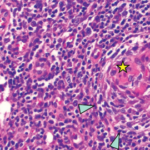
We report a case of a 27-year-old woman who was initially diagnosed with systemic lupus erythematosus (SLE), had features of scleroderma and was subsequently found to have lymph node biopsy consistent with multicentric Castleman disease (MCD). She also had serologic evidence of acute Epstein-Barr virus (EBV) infection (vs. reactivation of EBV). The occurrence of MCD in association with SLE and scleroderma is rare, and the relationship between these diseases and EBV remain areas of ongoing research. We report this case to increase awareness of this potential diagnostic and therapeutic dilemma.

Case

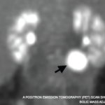
The patient is a 27-year-old African Caribbean woman who initially presented with malaise, dry cough, weight loss of 25 lbs. over four months, diffuse arthralgia, myalgia, hair loss, diaphoresis, numbness of extremities, bilateral ankle swelling, cold intolerance and Raynaud’s phenomenon. She was febrile, temperature 101.8º F, blood pressure of 102/57 mmHg, pulse of 111 bpm and respiratory rate of 24 breaths per minute. On physical exam she was cachectic, with tightness around the mouth, telangiectasia over the nasal bridge, digital ulcer on the left third digit, soft abdomen with hepatosplenomegaly, pitting edema of the lower extremities, bilateral axillary and femoral lymphadenopathy.

ad goes here:advert-1
ADVERTISEMENT
SCROLL TO CONTINUE

Labs were significant for hemoglobin (Hb) of 5.6 g/dL (normal range: 12–16 g/dL), a reticulocyte count of 3.11% (normal range: 0.5–2.9%), LDH 664 U/L (normal range: 135–214 U/L), haptoglobin of 3 mg/dL (normal range: 30–200 mg/dL), ferritin 589.7 ng/mL (normal range: 10–291 ng/mL), direct Coombs test positive for nonspecific cold agglutinin, an unremarkable peripheral blood smear and an ESR of 129 mm/hr (normal range: 0–20 mm/hr). She had an albumin of 2.6 g/dL (normal range: 2.8–5.7 g/dL) and 2+ proteinuria on dipstick. RF, ANA and c-ANCA were negative, but p-ANCA was positive. C3 was 45 mg/dL (normal range: 86–184 mg/dL) and C4 was 7 mg/dL (normal range: 20–58 mg/dL). HIV, human T cell lymphoma virus 1 (HTLV 1), hepatitis B and C screening, RPR, PPD and blood cultures were negative.

A non-contrast CT scan of the chest revealed bilateral axillary lymphadenopathy measuring up to 1.5 cm, trace bilateral pleural effusions and small pericardial effusion, but no consolidation. A CT scan of the abdomen with contrast revealed bulky retroperitoneal and pelvic lymphadenopathy with bulky external iliac nodes, the largest of which was 4.4 x 1.7 cm, ascites, hepatosplenomegaly, periportal edema, perihepatic ascites and pericholecystic fluid. 2D echo revealed an ejection fraction of 55–60%, mild tricuspid regurgitation, estimated pulmonary arterial pressure of 40 mmHg and a small to moderate pericardial effusion.

ad goes here:advert-2
ADVERTISEMENT
SCROLL TO CONTINUE

Page: 1 2 3 4 5 | Single Page
Share: 

Filed under:ConditionsSystemic Sclerosis Tagged with:Castleman's diseaseClinicalLupusoutcomepatient carerheumatologySclerodermaSLE

Related Articles
    A positron emission tomography (PET) scan shows a 3 cm, hypermetabolic mass adjacent to the stomach.

    Case Report: Elevated Inflammatory Markers & a Hypermetabolic Mass

    December 16, 2021

    Consulting rheumatologists often assess patients with atypical clinical presentations for the possibility of an underlying rheumatic disease. Inflammatory syndromes that are not clearly rheumatic in nature can be particularly challenging to diagnose. Here, we share the case of a young woman with a long-standing undiagnosed illness and highly elevated inflammatory markers, and describe the evaluation…

    Regressed germinal center with follicular dendritic cell prominence (star), onion-skinning (blue arrowhead) and increased vascularity (orange arrows) are seen.

    Case Report: Is It Castleman Disease, or Castleman-Like?

    October 13, 2021

    The difference between Castle­man disease and Castleman-like disease may be subtle, but it comes with significant ramifications. Case Presentation This case involves a pregnant 19-year-old woman who presents over multiple hospitalizations with concerns for systemic lupus erythematosus and macrophage activation syndrome. ad goes here:advert-1ADVERTISEMENTSCROLL TO CONTINUEAt 36 weeks’ gestation, the patient’s weight had dropped from…

    Search for Infectious Triggers of Rheumatic Disease Could Yield Options to Block Process

    January 19, 2016

    SAN FRANCISCO—Two hundred years ago, physicians knew nothing about what caused various forms of arthritis. Today, we have more clues about what may trigger rheumatic diseases, but still can’t pinpoint the culprits that set scores of inflammatory conditions in motion. Researchers discussed some of the background and recent findings that point to what immunologists call…

    What’s the Role of Epstein-Barr Virus Reactivation in Lupus Development?

    November 12, 2020

    A strong association between Epstein-Barr virus (EBV) infections and systemic lupus erythematosus (SLE) has been known since the 1970s. Although the etiology of SLE is not fully established, multiple genetic factors and polymorphisms in genes involved in the immune system have been implicated, with environmental factors also contributing to the development of this complex condition….

  • About Us
  • Meet the Editors
  • Issue Archives
  • Contribute
  • Advertise
  • Contact Us
  • Copyright © 2025 by John Wiley & Sons, Inc. All rights reserved, including rights for text and data mining and training of artificial technologies or similar technologies. ISSN 1931-3268 (print). ISSN 1931-3209 (online).
  • DEI Statement
  • Privacy Policy
  • Terms of Use
  • Cookie Preferences