She reported that the knee pain began after she performed some repairs on her automobile. One week prior to the rheumatology appointment, she saw an outside physician who believed she was experiencing an RA flare and prescribed a prednisone taper, without benefit. The knee pain progressed to include the right hip and posterior thigh, and she noticed swelling of the right lower extremity in the days prior to the appointment. She denied any trauma, recent travel or exposures. She reported a family history of unprovoked deep vein thrombosis (DVT) and pulmonary embolism in her mother that occurred at age 70. She denied a history of tobacco use.
Vital sign measurements were unremarkable, without fever, tachycardia or tachypnea. On physical examination, there was no evidence of synovitis in her upper extremity joints. The right lower extremity was mildly edematous compared with the left lower extremity. Warmth was noted unilaterally on the right lower extremity. Flexion and external rotation of the right hip joint elicited pain. No erythema, trauma or rash was noted on either extremity.
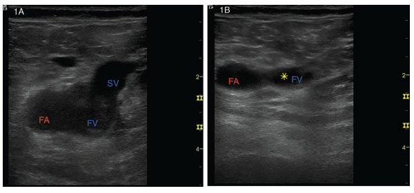
Figures 1A and B: (Left) A transverse view of the right common femoral artery (FA), right common femoral vein (FV) and right saphenous vein (SV). (Right) With gentle probe pressure, the saphenous vein collapses easily, but the common femoral vein is noncompressible. The yellow star represents the clot preventing collapse.
Due to concern for DVT, a two-region point-of-care ultrasound (POCUS) protocol was performed in the rheumatology clinic to scan for DVT of the right lower extremity. Lack of complete compressibility of the right femoral vein at the saphenous junction (see Figures 1A and B, above right) and the popliteal vein (see Figures 2A and B) was noted with intraluminal clot visualized. Color Flow Doppler was applied at the saphenous junction with disruption of flow due to the clot (see Figure 3). The left deep veins were fully compressible (see Figures 4A and B).
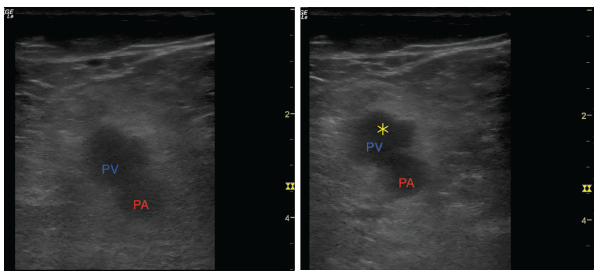
Figures 2A and B: (Left) A transverse view of the popliteal artery (PA) and popliteal vein (PV). (Right) Light compression with transducer shows an inability to collapse the popliteal vein. The yellow star represents the clot preventing collapse.