Video: Every Case Tells a Story| Webinar: ACR/CHEST ILD Guidelines in Practice

An official publication of the ACR and the ARP serving rheumatologists and rheumatology professionals

  • Conditions
    • Axial Spondyloarthritis
    • Gout and Crystalline Arthritis
    • Myositis
    • Osteoarthritis and Bone Disorders
    • Pain Syndromes
    • Pediatric Conditions
    • Psoriatic Arthritis
    • Rheumatoid Arthritis
    • Sjögren’s Disease
    • Systemic Lupus Erythematosus
    • Systemic Sclerosis
    • Vasculitis
    • Other Rheumatic Conditions
  • FocusRheum
    • ANCA-Associated Vasculitis
    • Axial Spondyloarthritis
    • Gout
    • Psoriatic Arthritis
    • Rheumatoid Arthritis
    • Systemic Lupus Erythematosus
  • Guidance
    • Clinical Criteria/Guidelines
    • Ethics
    • Legal Updates
    • Legislation & Advocacy
    • Meeting Reports
      • ACR Convergence
      • Other ACR meetings
      • EULAR/Other
    • Research Rheum
  • Drug Updates
    • Analgesics
    • Biologics/DMARDs
  • Practice Support
    • Billing/Coding
    • EMRs
    • Facility
    • Insurance
    • QA/QI
    • Technology
    • Workforce
  • Opinion
    • Patient Perspective
    • Profiles
    • Rheuminations
      • Video
    • Speak Out Rheum
  • Career
    • ACR ExamRheum
    • Awards
    • Career Development
  • ACR
    • ACR Home
    • ACR Convergence
    • ACR Guidelines
    • Journals
      • ACR Open Rheumatology
      • Arthritis & Rheumatology
      • Arthritis Care & Research
    • From the College
    • Events/CME
    • President’s Perspective
  • Search

Sonographic Diagnosis of Knuckle Pads

Pankaj Bansal, MD, RhMSUS, Eugene Kissin, MD, RhMSUS, & Fawad Aslam, MBBS, RhMSUS, RMSK  |  Issue: October 2022  |  October 10, 2022

The Case

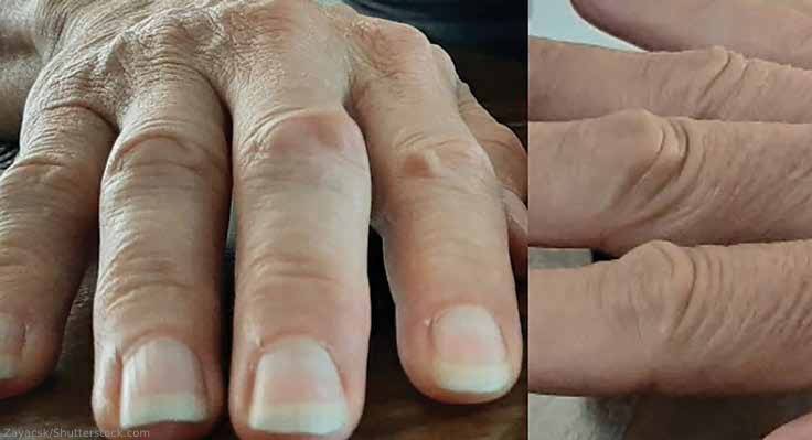
FIGURE 1: Knuckle pads over the proximal interphalangeal joints. (Click to enlarge.)

A 56-year-old white woman was evaluated for a one-year history of painless bumps on the dorsal aspect of the proximal interphalangeal (PIP) joints of both hands and suspected flexor tenosynovitis in her palms. On examination, small cystic nodules without erythema or tenderness were present on the dorsal aspect of several PIP joints (see Figure 1). The PIP joints had normal range of motion and no swelling or tenderness. She had induration along the course of the third flexor tendon without frank triggering or contractures. Hand radiographs were normal.

A musculoskeletal ultrasound examination was performed to assess for synovitis and tenosynovitis and to evaluate the nodules on the PIP joints and the palms. The ultrasound demonstrated the underlying third PIP joint and extensor tendon were normal, but it revealed a cystic, semi-lobulated lesion with hypoechoic and isoechoic features, which was partially compressible (see Figures 2 and 3) and exhibited a mild (grade 1) Doppler signal.

ad goes here:advert-1
ADVERTISEMENT
SCROLL TO CONTINUE

An evaluation of the palmar aspect of the right hand revealed an ill-defined hypoechoic lesion superficial to the flexor tendon proximal to the third metacarpophalangeal (MCP) joint (see Figures 4 and 5). It did not move with the underlying tendon, and the lesion had no Doppler signal. The underlying flexor tendon, the MCP joint and the A-1 pulley were normal. Diagnosis of PIP joint knuckle pads, in association with Dupuytren’s contracture, was made. No further intervention was recommended.

Discussion

Knuckle pads are benign subcutaneous nodules on the dorsal aspect of the PIP joints. Rarely, they are seen on the dorsal aspect of the MCP joints.1 These fibro-fatty nodules can be bilateral and are painless. Knuckle pads are usually idiopathic, although rare associations have been reported with repeated trauma and Dupuytren’s contractures.2-4 They are not associated with underlying joint or tendon abnormalities.

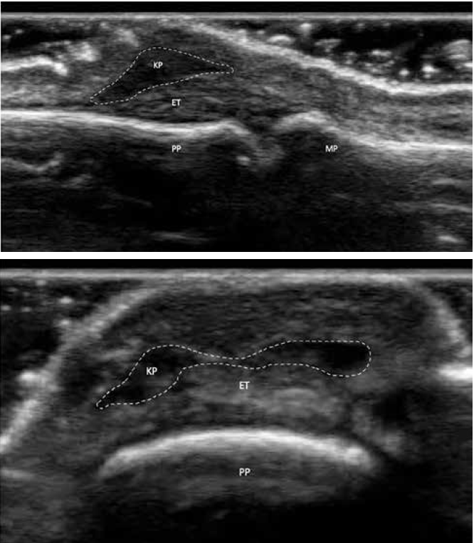
FIGURES 2 & 3: Dorsal longitudinal and transverse B-mode ultrasound views of the knuckle pads. Subcutaneous hypoechoic mass can be seen (enclosed in dotted lines). Key: MC: metacarpal; PP: proximal phalanx; ET: extensor tendon; KP: knuckle pad. (Click to enlarge).

No intervention is needed for these benign nodules. They usually come to the physician’s attention for cosmetic reasons or due to concerns of arthritis. An accurate diagnosis should be made after excluding other causes, such as gouty tophi, rheumatoid nodules, ganglion cysts, Bouchard nodes, synovitis and other rarer causes of nodules, such as multicentric reticulohistiocytosis.

Page: 1 2 3 4 | Single Page
Share: 

Filed under:Conditions Tagged with:case reportDupuytren's contractureknuckle padsUltrasound

Related Articles
    Musculoskeletal Ultrasound: A Valuable Tool for Diagnosing Rheumatic Illnesses

    Musculoskeletal Ultrasound: A Valuable Tool for Diagnosing Rheumatic Illnesses

    October 15, 2015

    Musculoskeletal (MSK) ultrasound is a valuable imaging modality for the practicing rheumatologist and provides an efficient tool with high diagnostic value in the evaluation of patients with musculoskeletal complaints. The use of MSK ultrasound has evolved in the U.S. due to the emergence of less-expensive, portable ultrasound units, which provide high-quality gray-scale and power Doppler…

    Using Ultrasound to Diagnose Carpal Tunnel Syndrome

    April 26, 2018

    Note: Updated May 2, 2018, to correct a link in the reference section. The error was introduced in editing. A 44-year-old Caucasian woman presented to the outpatient rheumatology clinic that had followed her for several years for rheumatoid arthritis. She was compliant with her regimen of hydroxychloroquine, etanercept and salsalate. Her chief complaint was worsening…

    Case Report: A Psoriatic Arthritis Patient with Dactylitis & Enthesitis

    September 20, 2018

    A 36-year-old woman presented at the Johns Hopkins Arthritis Center for a second opinion regarding a diagnosis of psoriatic arthritis (PsA). One year prior to our evaluation, she had developed pain and stiffness in her hands, feet, knees, ankles, elbows and shoulders. She had mild plaque psoriasis of the scalp and base of the neck,…

    Case Report: Perplexing Pulmonary Nodules

    March 14, 2022

    Pulmonary nodules are com­mon; most are benign, but the differential diagnosis is broad and includes life-threatening possibilities.1 Our patient is a former smoker who has a history of a complex autoimmune disease and multiple pulmonary nodules. This case was challenging, but clinical, radiographic and histologic clues helped lead to the correct diagnosis. Case Presentation The…

  • About Us
  • Meet the Editors
  • Issue Archives
  • Contribute
  • Advertise
  • Contact Us
  • Copyright © 2025 by John Wiley & Sons, Inc. All rights reserved, including rights for text and data mining and training of artificial technologies or similar technologies. ISSN 1931-3268 (print). ISSN 1931-3209 (online).
  • DEI Statement
  • Privacy Policy
  • Terms of Use
  • Cookie Preferences