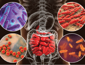
Bacteria (e.g., Bifidobacterium, Lactobacillus, Enterococcus and Escherichia coli) colonize different parts of digestive system.
Kateryna Kon / shutterstock.com
Evidence is accumulating that the microbiome may be an important part of the pathogenesis of many autoimmune diseases. Two recently published articles report on how translocation of the gut bacterium Enterococcus gallinarum drives autoimmunity in mice and humans, and on the role of other commensal bacteria in triggering immune responses—specifically to the autoantigen Ro60, which is targeted in systemic lupus erythematosus (SLE).1,2
“Both of these studies deal with the role of the microbes that live in our gut and other places of the human body of patients with autoimmune disease,” says Martin A. Kriegel, MD, PhD, adjunct professor of immunobiology at Yale University School of Medicine, New Haven, Conn., and lead author of the two reports. “The overarching theme in my laboratory is the role of the microbiome in autoimmune diseases. These results work toward a better understanding of how the microbiome fits into the development and treatment of these diseases.”
Indication of Bacterium Translocation
The study published in the March edition of Science looked at indicators of translocation of E. gallinarum in a hybrid autoimmune mouse model under pathogen-free conditions.1
The researchers studied the response in these mice to classic autoantigens in SLE as well as endogenous retrovirus glycoprotein 70, which had previously been shown to drive lupus kidney disease in this model. Mortality, lupus-related autoantibodies and autoimmune manifestations were all lowered after oral administration of antibiotics. In addition, translocation of the microbiota was successfully suppressed by vancomycin or ampicillin. Both also prevented mortality by autoimmune disease in this mouse model.

Dr. Kriegel
Uptake of an orally fed fluorescent dye into systemic circulation of the pathogen-free mice indicated impaired gut barrier function when compared with another mouse line that was not genetically predisposed to autoimmune disease. The researchers were able to detect bacterial growth in the mesenteric veins, mesenteric lymph nodes (MLN), spleen and liver, further indications of translocation.
When the group looked at full-length ribosomal DNA sequencing of single colonies from aerobic and anaerobic MLN, liver and spleen cultures, E. gallinarum predominated in all tissues including in the mesenteric veins of 82% of the lupus-prone mice. It was also visualized in situ by fluorescence within MLN and the liver. Control mice showed no bacterial growth.
Species-specific polymerase chain reaction (PCR) did not detect E. gallinarum DNA in stool samples of human or murine autoimmune hosts. However fecal or mucosal cultures followed by species-specific PCR consistently showed the bacterium in the feces, small intestine and liver of the hybrid mice, suggesting that some disease-relevant gut microbes are not easily detectable in conventional stool studies because they hide within the body.