Video: Every Case Tells a Story| Webinar: ACR/CHEST ILD Guidelines in Practice

An official publication of the ACR and the ARP serving rheumatologists and rheumatology professionals

  • Conditions
    • Axial Spondyloarthritis
    • Gout and Crystalline Arthritis
    • Myositis
    • Osteoarthritis and Bone Disorders
    • Pain Syndromes
    • Pediatric Conditions
    • Psoriatic Arthritis
    • Rheumatoid Arthritis
    • Sjögren’s Disease
    • Systemic Lupus Erythematosus
    • Systemic Sclerosis
    • Vasculitis
    • Other Rheumatic Conditions
  • FocusRheum
    • ANCA-Associated Vasculitis
    • Axial Spondyloarthritis
    • Gout
    • Psoriatic Arthritis
    • Rheumatoid Arthritis
    • Systemic Lupus Erythematosus
  • Guidance
    • Clinical Criteria/Guidelines
    • Ethics
    • Legal Updates
    • Legislation & Advocacy
    • Meeting Reports
      • ACR Convergence
      • Other ACR meetings
      • EULAR/Other
    • Research Rheum
  • Drug Updates
    • Analgesics
    • Biologics/DMARDs
  • Practice Support
    • Billing/Coding
    • EMRs
    • Facility
    • Insurance
    • QA/QI
    • Technology
    • Workforce
  • Opinion
    • Patient Perspective
    • Profiles
    • Rheuminations
      • Video
    • Speak Out Rheum
  • Career
    • ACR ExamRheum
    • Awards
    • Career Development
  • ACR
    • ACR Home
    • ACR Convergence
    • ACR Guidelines
    • Journals
      • ACR Open Rheumatology
      • Arthritis & Rheumatology
      • Arthritis Care & Research
    • From the College
    • Events/CME
    • President’s Perspective
  • Search

The Diagnostic View: Our Findings

Sunita Paudyal, MD, & Laura B. Herpel, MD  |  Issue: May 2018  |  May 17, 2018

Click here for the case.

Discussion

Image 1 demonstrates two nodules in the right lung, one over the lateral costophrenic sulcus (2.4 x1.7 cm, not shown); and another in the middle lobe (3.1 x 2.6 cm), with lobulated and spiculated margins (red arrow). There was no lymphadenopathy or pleural effusion. Of note, a normal chest X-ray from four years prior was available.

The CT was concerning for a neoplasm, which was corroborated by a subsequent PET-CT, showing hypermetabolic activity; however, there was no evidence of metastases. Baseline labs showed mild leukocytosis and microcytic anemia. Bronchoscopy and bronchoalveolar lavage were also unremarkable. Both adalimumab and methotrexate were discontinued, and she was treated with oral prednisone. A CT-guided lung biopsy was negative for malignancy and IgG4 disease showing inflammatory cells. A CT chest two weeks later (prior to a planned repeat biopsy) demonstrated significant interval decrease in the nodules’ size (not shown), and a biopsy was not done.

ad goes here:advert-1
ADVERTISEMENT
SCROLL TO CONTINUE

In a follow-up CT chest eight months later (Image 2) the nodules appeared almost resolved (red arrow). The patient was later restarted on methotrexate 20 mg SQ weekly, as well as weekly abatacept injections, with good control of RA and without recurrence of lung nodules.

Rheumatologists must be aware of potential lung diseases caused by TNF inhibitors (TNFIs) besides infections. A number of noninfectious pulmonary side effects are becoming increasingly recognized. These reactions include pulmonary fibrosis, granulomatous lesions, pneumonitis, as well as pulmonary nodules.1,2

ad goes here:advert-2
ADVERTISEMENT
SCROLL TO CONTINUE

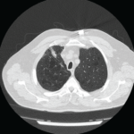
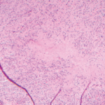
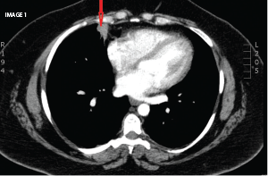
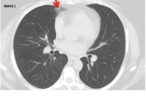
A study in the Journal of Rheumatology reported on RA patients receiving anti-tumor necrosis factor-alpha (TNF-alpha) therapy who developed pulmonary nodulosis or aseptic granulomatous lung disease. As in our patient, the nodules resolved after stopping TNFIs.2

The Diagnostic View: Assess Your Rheumatology Knowledge

Sunita Paudyal, MD, is an assistant professor in the Division of Rheumatology at the University of South Carolina School of Medicine. She completed her internal medicine residency at Providence Portland Medical Center, Oregon, and her fellowship at the Medical College of Georgia, Augusta.

Laura B. Herpel, MD, is board certified and fellowship trained in pulmonary medicine, critical care medicine and sleep medicine. Dr. Herpel earned her medical degree from the University of South Carolina School of Medicine. She completed her internal medicine residency and fellowship training at Johns Hopkins University School of Medicine in Baltimore.

References

  1. Thavarajah K, Wu P, Rhew E, et al. Pulmonary complications of tumor necrosis factor targeted therapy. Respir Med. 2009 May;103(5):661–669.
  2. Toussirot E, Berthelot JM, Pertuiset E, et al. pulmonary nodulosis and aseptic granulomatous lung disease occurring in patients with rheumatoid arthritis receiving tumor necrosis factor-alpha-blocking agent: A case series. J Rheumatol. 2009 Oct 1;36(11):2421–2427.

Page: 1 2 | Single Page
Share: 

Filed under:Conditions Tagged with:pulmonary nodulespulmonary nodulosisTNF inhibitors

Related Articles

    Case Report: Perplexing Pulmonary Nodules

    March 14, 2022

    Pulmonary nodules are com­mon; most are benign, but the differential diagnosis is broad and includes life-threatening possibilities.1 Our patient is a former smoker who has a history of a complex autoimmune disease and multiple pulmonary nodules. This case was challenging, but clinical, radiographic and histologic clues helped lead to the correct diagnosis. Case Presentation The…

    The Granulomatosis of Wegener’s

    May 16, 2011

    Delving deeper into the nonvasculitis aspects of the disease

    A Mystery Case of Abdominal Masses

    May 13, 2021

    Common variable immuno­deficiency (CVID) is a common primary immuno­deficiency disease, with an estimated incidence of one per 25,000–50,000 individuals.1 The classic presen­tation includes frequent bacterial infections, secondary to dysfunctional B cell differ­entiation, impaired immunoglobulin production and diminished antibody response. The clinical presentation may be heterogenous and may include granulomatous disease as an uncommon manifestation. Granulo­matous…

    Case Study in Dermatology: Tender Papules on Elbows, Hands in RA

    February 17, 2016

    The Case A 41-year-old woman was referred to the dermatology clinic for a three-month history of tender lesions on her elbows and around the joints of her hands. Her medical history was notable for seropositive, non-erosive rheumatoid arthritis (RA), which was being treated with 5 mg pred­nisone daily, 10 mg methotrexate weekly by mouth, 200…

  • About Us
  • Meet the Editors
  • Issue Archives
  • Contribute
  • Advertise
  • Contact Us
  • Copyright © 2025 by John Wiley & Sons, Inc. All rights reserved, including rights for text and data mining and training of artificial technologies or similar technologies. ISSN 1931-3268 (print). ISSN 1931-3209 (online).
  • DEI Statement
  • Privacy Policy
  • Terms of Use
  • Cookie Preferences