Video: Every Case Tells a Story| Webinar: ACR/CHEST ILD Guidelines in Practice

An official publication of the ACR and the ARP serving rheumatologists and rheumatology professionals

  • Conditions
    • Axial Spondyloarthritis
    • Gout and Crystalline Arthritis
    • Myositis
    • Osteoarthritis and Bone Disorders
    • Pain Syndromes
    • Pediatric Conditions
    • Psoriatic Arthritis
    • Rheumatoid Arthritis
    • Sjögren’s Disease
    • Systemic Lupus Erythematosus
    • Systemic Sclerosis
    • Vasculitis
    • Other Rheumatic Conditions
  • FocusRheum
    • ANCA-Associated Vasculitis
    • Axial Spondyloarthritis
    • Gout
    • Psoriatic Arthritis
    • Rheumatoid Arthritis
    • Systemic Lupus Erythematosus
  • Guidance
    • Clinical Criteria/Guidelines
    • Ethics
    • Legal Updates
    • Legislation & Advocacy
    • Meeting Reports
      • ACR Convergence
      • Other ACR meetings
      • EULAR/Other
    • Research Rheum
  • Drug Updates
    • Analgesics
    • Biologics/DMARDs
  • Practice Support
    • Billing/Coding
    • EMRs
    • Facility
    • Insurance
    • QA/QI
    • Technology
    • Workforce
  • Opinion
    • Patient Perspective
    • Profiles
    • Rheuminations
      • Video
    • Speak Out Rheum
  • Career
    • ACR ExamRheum
    • Awards
    • Career Development
  • ACR
    • ACR Home
    • ACR Convergence
    • ACR Guidelines
    • Journals
      • ACR Open Rheumatology
      • Arthritis & Rheumatology
      • Arthritis Care & Research
    • From the College
    • Events/CME
    • President’s Perspective
  • Search

The Power of Power Doppler: Ultrasound Imaging in RA

Jason Liebowitz, MD, FACR  |  Issue: April 2022  |  March 23, 2022

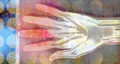
BALTIMORE—Rheumatology is a field in which all data points are considered in building the case for a patient’s diagnosis and seeking to clarify disease activity and prognosis. In recent years, information gleaned through ultrasound imaging has helped supplement the data gathered from the traditional history, physical exam, and laboratory and radiographic studies alone.

At the 17th Annual Advances in the Diagnosis and Treatment of the Rheumatic Diseases meeting at Johns Hopkins University School of Medicine, Baltimore, Dana DiRenzo, MD, MHS, RhMSUS, instructor of medicine, Johns Hopkins University School of Medicine, provided an extremely helpful overview of the use of ultrasound imaging in patients with inflammatory arthritis.

ad goes here:advert-1
ADVERTISEMENT
SCROLL TO CONTINUE

Limitations of Traditional Examinations

Dr. DiRenzo began by noting some of the limitations of the traditional clinical examination in evaluating patients for rheumatoid arthritis (RA). Disease activity scores can sometimes be artificially elevated in patients with comorbidities that increase the tender joint count even in the absence of frank joint swelling, she explained. The issue of inter-observer variance of clinical joint assessments also arises, with different clinicians having different assessments of the presence or absence of synovitis in the same joint for a specific patient. More recently, a newfound appreciation has developed for the entity of “subclinical synovitis,” which may not be easily detectable on the clinical exam.

For the definition of terms for ultrasound findings, Dr. DiRenzo referenced the Outcome Measures in Rheumatology (OMERACT) group definitions for ultrasound-detected pathologies.

ad goes here:advert-2
ADVERTISEMENT
SCROLL TO CONTINUE

Synovitis is defined as the presence of a hypoechoic synovial hypertrophy regardless of the presence of effusion or any grade of Doppler signal.

The term erosion is defined as intra- and/or extra-articular discontinuity of bone surface that is visible in two perpendicular planes.1

Other helpful terms include gray scale, which is the modality used to detect lesions of synovial effusion and hypertrophy, and power Doppler, which is the modality used to detect blood flow to the synovial membrane.2

Baseline Ultrasound Assessment

Ultrasound is used in rheumatology for diagnosis and disease monitoring. It can also assist in predicting prognosis, Dr. DiRenzo noted. With regard to diagnosis, Horton et al. evaluated 60 patients with new-onset undifferentiated arthritis who did not meet 2010 ACR/European League Against Rheumatism (EULAR) RA classification criteria.3 These patients underwent baseline ultrasound assessment and then were followed prospectively, with 22% ultimately meeting criteria for RA, and 53% (32 of the 60 patients) receiving methotrexate within 12 months after enrollment. The authors found that severity of gray scale synovitis detected across 26 joints was predictive of progression to RA and of methotrexate use, independent of the clinical swollen joint count or disease activity score.

Page: 1 2 3 | Single Page
Share: 

Filed under:ConditionsMeeting ReportsRheumatoid Arthritis Tagged with:Advances in the Diagnosis and Treatment of the Rheumatic Diseases meetingdiagnostic imagingimagingRheumatoid Arthritis (RA)Ultrasound

Related Articles
    Musculoskeletal Ultrasound: A Valuable Tool for Diagnosing Rheumatic Illnesses

    Musculoskeletal Ultrasound: A Valuable Tool for Diagnosing Rheumatic Illnesses

    October 15, 2015

    Musculoskeletal (MSK) ultrasound is a valuable imaging modality for the practicing rheumatologist and provides an efficient tool with high diagnostic value in the evaluation of patients with musculoskeletal complaints. The use of MSK ultrasound has evolved in the U.S. due to the emergence of less-expensive, portable ultrasound units, which provide high-quality gray-scale and power Doppler…

    The Heterogeneity of Psoriatic Arthritis

    November 21, 2023

    SAN DIEGO—Differences between psoriatic arthritis and rheumatoid arthritis highlight the need for the development of imaging modalities, laboratory tests and other biomarkers that are explored and validated specifically for PsA to advance the goal of personalized or precision medicine. In this article, expert David S. Pisetsky, MD, PhD, explores the top research in psoriatic arthritis presented at ACR Convergence 2023.

    Tips for Implementing Ultrasound Training in Rheumatology Fellowships

    February 13, 2020

    ATLANTA—Point-of-care ultrasound education mainly has occurred at the undergraduate level at U.S. medical schools, but rheumatology fellowship training programs are rapidly catching up and integrating it into their curricula, according to two program directors who reviewed the state of rheumatology ultrasound education, including potential barriers to its implementation, on Nov. 12 at the 2019 ACR/ARP…

    Target Remission

    March 1, 2007

    Strategies to identify and track remission in your RA patients

  • About Us
  • Meet the Editors
  • Issue Archives
  • Contribute
  • Advertise
  • Contact Us
  • Copyright © 2025 by John Wiley & Sons, Inc. All rights reserved, including rights for text and data mining and training of artificial technologies or similar technologies. ISSN 1931-3268 (print). ISSN 1931-3209 (online).
  • DEI Statement
  • Privacy Policy
  • Terms of Use
  • Cookie Preferences