Video: Every Case Tells a Story| Webinar: ACR/CHEST ILD Guidelines in Practice

An official publication of the ACR and the ARP serving rheumatologists and rheumatology professionals

  • Conditions
    • Axial Spondyloarthritis
    • Gout and Crystalline Arthritis
    • Myositis
    • Osteoarthritis and Bone Disorders
    • Pain Syndromes
    • Pediatric Conditions
    • Psoriatic Arthritis
    • Rheumatoid Arthritis
    • Sjögren’s Disease
    • Systemic Lupus Erythematosus
    • Systemic Sclerosis
    • Vasculitis
    • Other Rheumatic Conditions
  • FocusRheum
    • ANCA-Associated Vasculitis
    • Axial Spondyloarthritis
    • Gout
    • Psoriatic Arthritis
    • Rheumatoid Arthritis
    • Systemic Lupus Erythematosus
  • Guidance
    • Clinical Criteria/Guidelines
    • Ethics
    • Legal Updates
    • Legislation & Advocacy
    • Meeting Reports
      • ACR Convergence
      • Other ACR meetings
      • EULAR/Other
    • Research Rheum
  • Drug Updates
    • Analgesics
    • Biologics/DMARDs
  • Practice Support
    • Billing/Coding
    • EMRs
    • Facility
    • Insurance
    • QA/QI
    • Technology
    • Workforce
  • Opinion
    • Patient Perspective
    • Profiles
    • Rheuminations
      • Video
    • Speak Out Rheum
  • Career
    • ACR ExamRheum
    • Awards
    • Career Development
  • ACR
    • ACR Home
    • ACR Convergence
    • ACR Guidelines
    • Journals
      • ACR Open Rheumatology
      • Arthritis & Rheumatology
      • Arthritis Care & Research
    • From the College
    • Events/CME
    • President’s Perspective
  • Search

Tips from a Cardiothoracic Radiologist: Interstitial Lung Disease & Radiology for the Rheumatologist

Samantha C. Shapiro, MD  |  Issue: July 2022  |  June 17, 2022

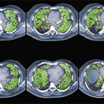
First, assess the distribution of abnormalities. What area of the lung is involved? Are the abnormalities located at the top or bottom (i.e., apical or basilar)? Inner or outer (i.e., central or peripheral)?

Next, describe the dominant pattern of the abnormalities. Look for the following:

ad goes here:advert-1
ADVERTISEMENT
SCROLL TO CONTINUE
  • Reticulation: too many fine lines;
  • Groundglass: an increase in lung tissue density that doesn’t obscure the vessels;
  • Consolidation: an increased density obscures the vessels and air bronchograms may be present;
  • Nodules: too many dots;
  • Honeycombing: small cystic black spaces touching each other, a sign of end-stage disease;
  • Air trapping on expiratory images; and
  • Traction bronchiectasis: dilated airways in areas of abnormal lung, another sign of fibrosis.

“Description and distribution may be diagnostic, obviating the need for lung biopsy,” Dr. Guttentag said. “They also help inform therapeutic intervention and prognosis. For example, a patient with honeycombing and traction bronchiectasis has established fibrosis that won’t respond to immunosuppressive therapy. But groundglass and consolidation could indicate active inflammation that’s potentially reversible.”

Most importantly, consider the clinical information. “Do the CT findings support your clinical diagnosis?” Dr. Guttentag asked. “The age and sex of the patient, acuity of symptoms and social and occupational history are all relevant here. And of course, is there clinical evidence of connective tissue disease [CTD]?”

ad goes here:advert-2
ADVERTISEMENT
SCROLL TO CONTINUE

‘Don’t ever order a CT chest with & without contrast unless you’ve spoken to radiology & there’s a good reason for it. It just doubles the radiation dose.’ —Dr. Guttentag

CTD ILD

Generally, CTD presents in the lungs as one of various patterns of idiopathic interstitial pneumonia (IIP). Dr. Guttentag said, “Think of IIP as the way the lung is reacting to an insult. These are nonspecific histologic patterns of lung injury. The lung has limited ways of responding and trying to heal, and these insults may be autoimmune, inhalational, infectious, idiopathic or drug induced. The history, clinical exam and serologies are going to help put the pattern in the context of a clinical picture.”

ILD may precede clinical manifestations of CTD by months to years. “Up to 15% of cases of IIP will eventually be diagnosed with CTD, but IIP patterns aren’t specific to one CTD. There’s a lot of overlap,” Dr. Guttentag said.

“For example, many CTDs present with nonspecific interstitial pneumonia (NSIP). It’s ‘nonspecific.’ On the other hand, the usual interstitial pneumonia (UIP) pattern is most commonly seen in patients with rheumatoid lung. Systemic lupus erythematosus tends to show up with pleural-pericardial disease or hemorrhage. And systemic sclerosis almost always manifests with NSIP. So if the pattern doesn’t fit with what you typically see in that disease, consider whether they have something else superimposed. History, lab data and communication with radiology are critical.”1,2

Page: 1 2 3 | Single Page
Share: 

Filed under:ConditionsMeeting ReportsOther ACR meetings Tagged with:ACR Education Exchangecomputed tomographyconnective tissue diseaseCT scanCTD-ILDILDinterstitial lung disease (ILD)lungsRadiologistVasculitis

Related Articles

    What Rheumatologists Need to Know about Diagnosing and Managing Interstitial Lung Disease (ILD)

    December 1, 2012

    Patients with systemic sclerosis (SSc), poly-/dermatomyositis (PM/DM), or rheumatoid arthritis (RA) appear to carry the greatest risk for developing connective tissue disease-associated interstitial lung disease (CTD-ILD)
     

    Scleroderma & ILD: Practical Tips on the Diagnosis & Management of Systemic Sclerosis-Associated Interstitial Lung Disease

    June 15, 2022

    No one-size-fits-all approach exists for the care and treatment of patients with systemic sclerosis (SSc) and SSc with pulmonary involvement. Here, experts discuss some best clinical practices for these patients.

    When Interstitial Lung Disease and Connective Tissue Disorder Intersect in Rheumatology Patients

    February 15, 2017

    SAN FRANCISCO—Interstitial lung disease (ILD) represents a heterogeneous group of disorders characterized by inflammation or fibrosis of the lungs. The disorders are also associated with a spectrum of connective tissue diseases (CTDs). ILD is a common manifestation of CTDs, such as scleroderma, poly-/dermatomyositis and rheumatoid arthritis—and is a leading cause of morbidity and mortality in…

    Tocilizumab Effective for Early SSc-Associated Interstitial Lung Disease

    September 22, 2021

    Treatment with tocilizimab preserved lung function in patients with systemic sclerosis (SSc) and interstitial lung disease (ILD) regardless of a patient’s level of lung involvement, according to a recent study.

  • About Us
  • Meet the Editors
  • Issue Archives
  • Contribute
  • Advertise
  • Contact Us
  • Copyright © 2025 by John Wiley & Sons, Inc. All rights reserved, including rights for text and data mining and training of artificial technologies or similar technologies. ISSN 1931-3268 (print). ISSN 1931-3209 (online).
  • DEI Statement
  • Privacy Policy
  • Terms of Use
  • Cookie Preferences