Video: Every Case Tells a Story| Webinar: ACR/CHEST ILD Guidelines in Practice

An official publication of the ACR and the ARP serving rheumatologists and rheumatology professionals

  • Conditions
    • Axial Spondyloarthritis
    • Gout and Crystalline Arthritis
    • Myositis
    • Osteoarthritis and Bone Disorders
    • Pain Syndromes
    • Pediatric Conditions
    • Psoriatic Arthritis
    • Rheumatoid Arthritis
    • Sjögren’s Disease
    • Systemic Lupus Erythematosus
    • Systemic Sclerosis
    • Vasculitis
    • Other Rheumatic Conditions
  • FocusRheum
    • ANCA-Associated Vasculitis
    • Axial Spondyloarthritis
    • Gout
    • Psoriatic Arthritis
    • Rheumatoid Arthritis
    • Systemic Lupus Erythematosus
  • Guidance
    • Clinical Criteria/Guidelines
    • Ethics
    • Legal Updates
    • Legislation & Advocacy
    • Meeting Reports
      • ACR Convergence
      • Other ACR meetings
      • EULAR/Other
    • Research Rheum
  • Drug Updates
    • Analgesics
    • Biologics/DMARDs
  • Practice Support
    • Billing/Coding
    • EMRs
    • Facility
    • Insurance
    • QA/QI
    • Technology
    • Workforce
  • Opinion
    • Patient Perspective
    • Profiles
    • Rheuminations
      • Video
    • Speak Out Rheum
  • Career
    • ACR ExamRheum
    • Awards
    • Career Development
  • ACR
    • ACR Home
    • ACR Convergence
    • ACR Guidelines
    • Journals
      • ACR Open Rheumatology
      • Arthritis & Rheumatology
      • Arthritis Care & Research
    • From the College
    • Events/CME
    • President’s Perspective
  • Search

Vasculitis

Gary S. Hoffman, MD  |  Issue: June 2009  |  June 1, 2009

These observations raise important questions about a fascinating animal model of aortitis initially studied more than 10 years ago.12,13 A usually nonpathogenic herpes virus (γHV68) was provided as a standard inoculum to immunologically intact mice, as well as IFNγ- and IFNγ−receptor(R)-deficient mice. While the intact mice did not develop illness, the IFNγ- and IFNγ−R-deficient mice died from aortic rupture or dissection at approximately eight to 10 weeks. When animals were serially sacrificed and studied from one to six weeks postinoculation, it was apparent that widespread viremia had occurred but that virus could be effectively cleared from the IFNγ- and IFNγ–R-deficient mice from all organs except the aortic root. Thus, despite their lack of either IFNγ- or IFNγ−R, the gene-deficient animals possessed adequate immunological redundancy in other organs and within the distal thoracic and abdominal aorta to clear virus. However, the aortic root was uniquely vulnerable.
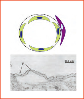
Figure 2B: Fenestrated EC arrangements are found in capillaries that allow rapid passage of large molecules such as in endocrine and exocrine glands, gastrointestinal mucosa, and intestinal villi. In the kidney, the renal glomeruli in which pericytes are mesangial cells, a fenestrated arrangement is present; however, this is not the case within the renal vasa recta where a continuous EC microanatomy is maintained.

Conclusions

Vessels are not merely anatomic conduits that carry blood, gases, nutrients, and waste products. Furthermore, they cannot be considered in simplistic terms based only on vessel size. It is clear that vessels, even of the same caliber from different organs, have important differences in their microanatomy, chemistry, and function. They work as partners in tissue development and have unique barrier and immunologic roles in different sites. Vessels, like all other tissues, also undergo changes in time. They can be sites of topographically selective chemical and other forms of injury or can selectively harbor infectious agents. The injury–repair process that is part of adapting to life’s challenges modifies substrate and is susceptible to errors in response and repair that may include mutations. Although this process plays an important role in the pathogenesis of cancer, it has not been examined within affected vessels in vasculitis. Might mutations provide neo-antigens as one means of stimulating an immune response against vessels, and could this response even be a functionally “normal” attempt to clear a “foreign” protein?

ad goes here:advert-1
ADVERTISEMENT
SCROLL TO CONTINUE

These questions do not diminish the importance of gender effects, immunological senescence, or inheritance in establishing disease predisposition. They do, however, emphasize that vessels are site-variable, complex, specialized organs, with qualities that are probably important in determining disease patterns. We can ill afford to ignore the unique properties of vascular territories and how they change over time if we wish to better understand the pathogenesis of the vasculitides. We should begin to apply lessons from embryology and vascular biology and reach beyond the simplistic notion of vasculitis being “understood” based principally on vessel size.

Page: 1 2 3 4 5 6 | Single Page
Share: 

Filed under:ConditionsVasculitis Tagged with:DiagnosisEtiologyTreatmentVasculitis

Related Articles

    Fellow’s Forum Case Report: Takayasu’s Arteritis

    May 1, 2014

    How this rare form of large-vessel vasculitis affects different portions, branches of aorta and ways to diagnose, treat and manage the disease

    Updates on Giant Cell Arteritis

    March 19, 2018

    SAN DIEGO—Recent research tells us more about giant cell arteritis (GCA) to help rheumatologists more accurately diagnose and effectively treat patients with this type of vasculitis. On Nov. 6 at the ACR/ARHP Annual Meeting, three experts explored the latest findings on GCA pathogenesis, diagnostic approaches, imaging modalities and growing treatment options. GCA: What’s Really Happening?…

    Two Inflammatory Conditions—Polymyalgia Rheumatica and Giant Cell Arteritis—Share Clinical Connection

    March 1, 2013

    Polymyalgia rheumatica (PMR) and giant cell arteritis (GCA) have common clinical and epidemiologic links, but they need not occur synchronously

    crystal light / shutterstock.com

    Rheumatology Case Report: Hand Abnormalities Feature of Fetal Hydantoin Syndrome

    November 6, 2017

    Research has shown that anticonvulsants are teratogens and pose a risk for fetal malformations. Meadow was the first to note a possible link between congenital abnormalities and maternal use of anticonvulsive drug in 1968.1 In 1974, Barr et al noted hypoplasia and irregular ossification of the digital distal phalanges with nail dystrophy in children born to…

  • About Us
  • Meet the Editors
  • Issue Archives
  • Contribute
  • Advertise
  • Contact Us
  • Copyright © 2025 by John Wiley & Sons, Inc. All rights reserved, including rights for text and data mining and training of artificial technologies or similar technologies. ISSN 1931-3268 (print). ISSN 1931-3209 (online).
  • DEI Statement
  • Privacy Policy
  • Terms of Use
  • Cookie Preferences