ACR Convergence 2025| Video: Rheuminations on Milestones & Ageism

An official publication of the ACR and the ARP serving rheumatologists and rheumatology professionals

  • Conditions
    • Axial Spondyloarthritis
    • Gout and Crystalline Arthritis
    • Myositis
    • Osteoarthritis and Bone Disorders
    • Pain Syndromes
    • Pediatric Conditions
    • Psoriatic Arthritis
    • Rheumatoid Arthritis
    • Sjögren’s Disease
    • Systemic Lupus Erythematosus
    • Systemic Sclerosis
    • Vasculitis
    • Other Rheumatic Conditions
  • FocusRheum
    • ANCA-Associated Vasculitis
    • Axial Spondyloarthritis
    • Gout
    • Lupus Nephritis
    • Psoriatic Arthritis
    • Rheumatoid Arthritis
    • Sjögren’s Disease
    • Systemic Lupus Erythematosus
  • Guidance
    • Clinical Criteria/Guidelines
    • Ethics
    • Legal Updates
    • Legislation & Advocacy
    • Meeting Reports
      • ACR Convergence
      • Other ACR meetings
      • EULAR/Other
    • Research Rheum
  • Drug Updates
    • Analgesics
    • Biologics/DMARDs
  • Practice Support
    • Billing/Coding
    • EMRs
    • Facility
    • Insurance
    • Technology
      • Information Technology
      • Apps
    • QA/QI
    • Workforce
  • Opinion
    • Patient Perspective
    • Profiles
    • Rheuminations
      • Video
    • Speak Out Rheum
  • Career
    • ACR ExamRheum
    • Awards
    • Career Development
      • Education & Training
    • Certification
  • ACR
    • ACR Home
    • ACR Convergence
    • ACR Guidelines
    • Journals
      • ACR Open Rheumatology
      • Arthritis & Rheumatology
      • Arthritis Care & Research
    • From the College
    • Events/CME
    • President’s Perspective
  • Search

What Clinicians Should Know About Midfoot Osteoarthritis

Yvonne M. Golightly, PT, MS, PhD, Jill Halstead, BSc, MSc, PhD, MRCPod, John B. Arnold, PhD, Lara S. Chapman, MSc, BPod, Catherine J. Bowen, PhD, BPod, Marian T. Hannan, DSc, MPH, Hylton B. Menz, PhD, BPod, Kade L. Paterson, PhD, BPod, BAppSci(Hons) & Martin J. Thomas, PhD, MCSP  |  Issue: October 2025  |  October 8, 2025

Accessing appropriate foot care is often difficult, especially in underserved communities, due to long wait times, limited availability of foot health care and financial barriers that may delay diagnosis and treatment.12,13

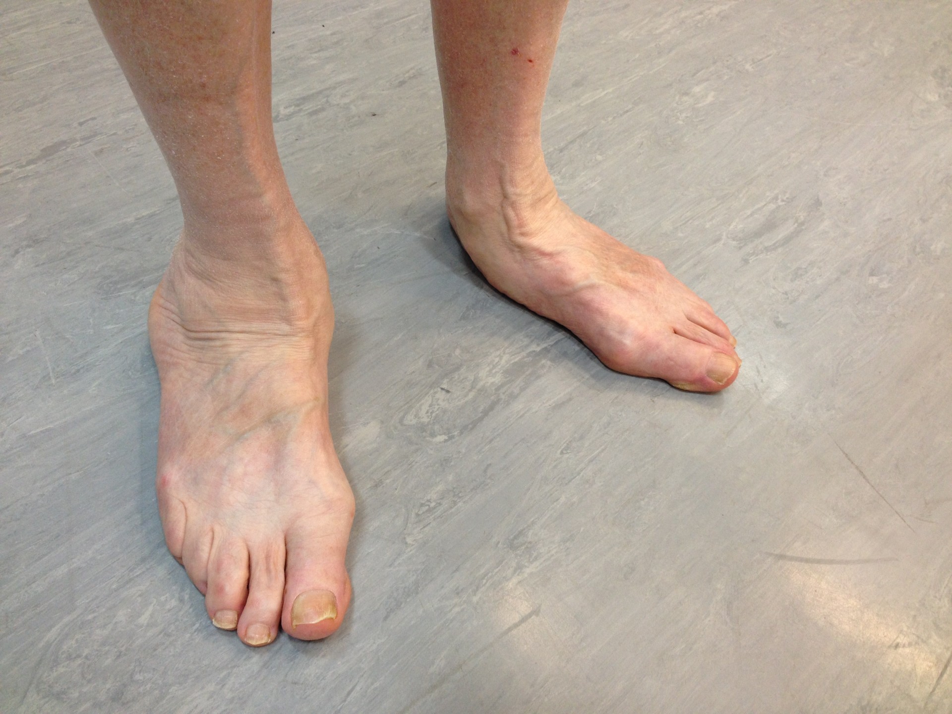
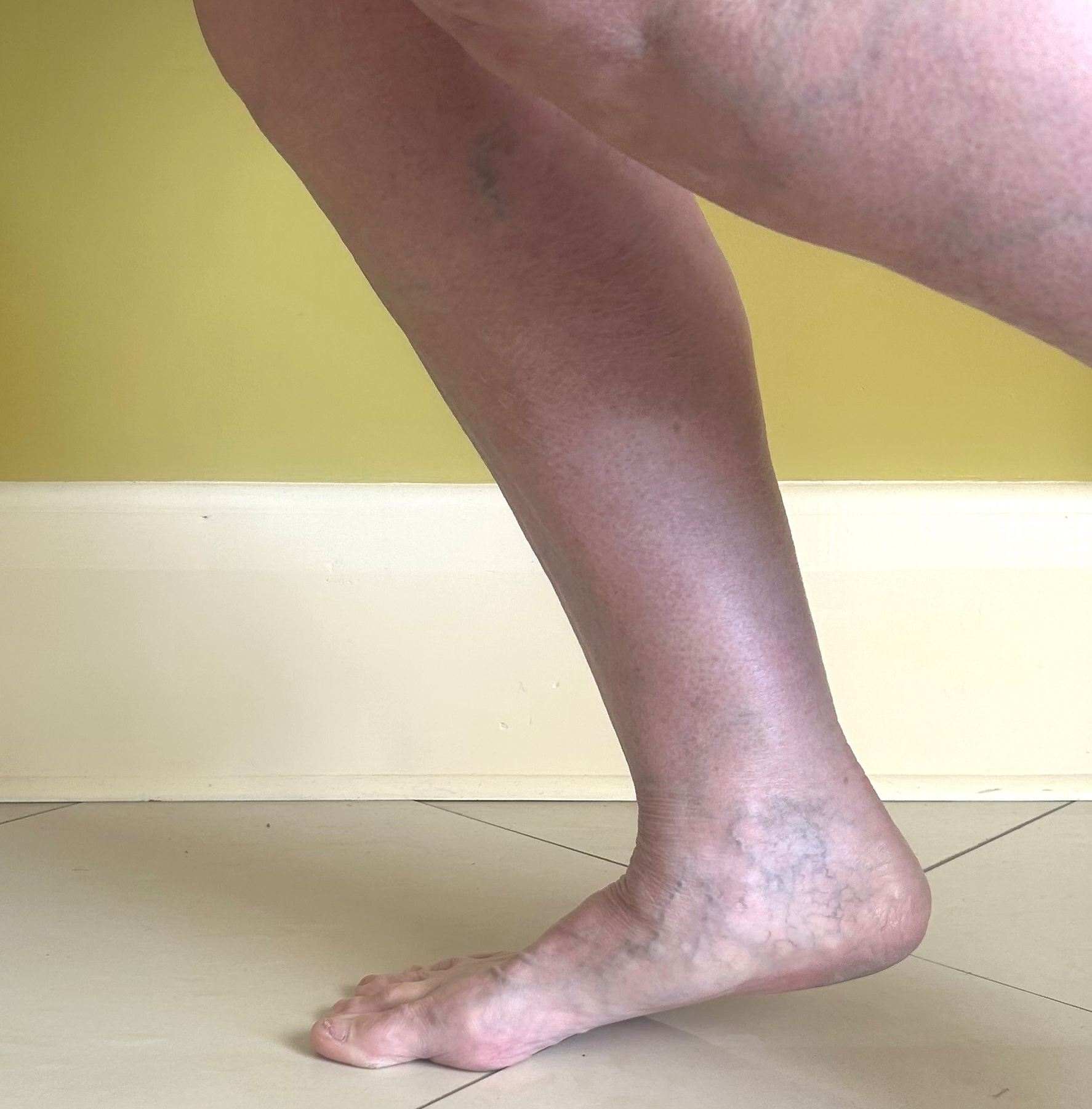
FIGURES 2: Midfoot OA and flat feet (left), and midfoot flattening during heel raise (right). (Click to enlarge.)

Disparities in research amplify clinical bias, with under-representation across groups leaving gaps in knowledge and care.14 Certain populations often face systemic barriers, such as limited insurance coverage, transportation issues, and fewer culturally competent providers.15 Addressing these inequalities is essential for improving care and QoL for all people living with midfoot OA.

ad goes here:advert-1
ADVERTISEMENT
SCROLL TO CONTINUE

How Is It Evaluated Clinically?

Imaging

Radiographic diagnosis of midfoot OA has traditionally utilized the Kellgren-Lawrence system.16 More recently, the La Trobe Foot Atlas was developed to identify radiographic OA across four joints (medial and intermediate cuneiform-metatarsal joints, talon­avicular joint and navicular-first cuneiform joint).17

This atlas incorporates cardinal features of OA—osteophytes and joint space narrowing—allowing for four levels of severity and diagnosis of radiographic changes (scores ≥2) plus symptoms in the corresponding region. Magnetic resonance imaging (MRI), although less accessible than X-ray, may also be used with a semiquantitative scoring system that includes all joints of the midfoot.18

ad goes here:advert-2
ADVERTISEMENT
SCROLL TO CONTINUE

Physical Exam

Midfoot OA commonly presents as persistent dorsal midfoot pain, worsening with weight-bearing and high-stress activities on the midfoot (e.g., rising onto the ball of foot, walking up or down stairs). Tight footwear pressing on the top of the midfoot may also be painful.

Pain may be reproduced with joint palpation or movement, but the prox­imity of the joints can make anatomical localization and diagnosis challenging without imaging.

Tarsometatarsal (Lisfranc) involvement may be identified by stabilizing the hind foot and plantarflexing the respective metatarsal (i.e., piano key test), although diagnostic accuracy is not established.19 Dorsal osteophytes may be palpable in advanced cases but limited in earlier midfoot OA.20 Localized midfoot joint swelling may be present and this is best detected with ultrasound examination.21 For unilateral symptoms, comparison to the other foot may be useful.

Midfoot pain after an axial load to a plantarflexed foot (e.g., a stumble) or following direct blunt trauma should raise suspicion of Lisfranc ligament injury or fracture, warranting urgent imaging.

If a patient has night pain, neurological symptoms, persistent inflammation and swelling or signs of infection, more serious conditions should be considered and urgent investigation undertaken.

Page: 1 2 3 4 5 | Single Page
Share: 

Filed under:ConditionsGuidanceInterprofessional PerspectiveOsteoarthritis and Bone DisordersResearch Rheum Tagged with:DisparitiesExercisefootimagingmidfoot oteoarthritisNSAIDsorthoticsPainpodiatryquality of life

Related Articles

    Foot Pain a Burden for Rheumatology Patients

    January 13, 2012

    Researchers say it may even correlate with back, hip, and knee pain

    Changing Our Thinking on Osteoarthritis

    March 1, 2010

    It’s time we changed our thinking on osteoarthritis

    Standing on a Hidden Burden: The Oft-Overlooked Problem of Foot and Ankle Osteoarthritis

    Standing on a Hidden Burden: The Oft-Overlooked Problem of Foot and Ankle Osteoarthritis

    November 14, 2021

    The high prevalence and significant burden of osteoarthritis (OA) are well known. But it’s becoming apparent that OA research efforts don’t necessarily match disease prevalence or disability. The International Foot and Ankle Osteoarthritis Consortium (IFOAC) is an international organization of volunteers that seeks to highlight a major and often overlooked problem in OA research—foot and…

    Osteoarthritis Quo Vadis

    March 1, 2008

    Our knowledge of OA has progressed far—does a cure lie ahead?

  • About Us
  • Meet the Editors
  • Issue Archives
  • Contribute
  • Advertise
  • Contact Us
  • Copyright © 2025 by John Wiley & Sons, Inc. All rights reserved, including rights for text and data mining and training of artificial technologies or similar technologies. ISSN 1931-3268 (print). ISSN 1931-3209 (online).
  • DEI Statement
  • Privacy Policy
  • Terms of Use
  • Cookie Preferences