Video: Every Case Tells a Story| Webinar: ACR/CHEST ILD Guidelines in Practice

An official publication of the ACR and the ARP serving rheumatologists and rheumatology professionals

  • Conditions
    • Axial Spondyloarthritis
    • Gout and Crystalline Arthritis
    • Myositis
    • Osteoarthritis and Bone Disorders
    • Pain Syndromes
    • Pediatric Conditions
    • Psoriatic Arthritis
    • Rheumatoid Arthritis
    • Sjögren’s Disease
    • Systemic Lupus Erythematosus
    • Systemic Sclerosis
    • Vasculitis
    • Other Rheumatic Conditions
  • FocusRheum
    • ANCA-Associated Vasculitis
    • Axial Spondyloarthritis
    • Gout
    • Psoriatic Arthritis
    • Rheumatoid Arthritis
    • Systemic Lupus Erythematosus
  • Guidance
    • Clinical Criteria/Guidelines
    • Ethics
    • Legal Updates
    • Legislation & Advocacy
    • Meeting Reports
      • ACR Convergence
      • Other ACR meetings
      • EULAR/Other
    • Research Rheum
  • Drug Updates
    • Analgesics
    • Biologics/DMARDs
  • Practice Support
    • Billing/Coding
    • EMRs
    • Facility
    • Insurance
    • QA/QI
    • Technology
    • Workforce
  • Opinion
    • Patient Perspective
    • Profiles
    • Rheuminations
      • Video
    • Speak Out Rheum
  • Career
    • ACR ExamRheum
    • Awards
    • Career Development
  • ACR
    • ACR Home
    • ACR Convergence
    • ACR Guidelines
    • Journals
      • ACR Open Rheumatology
      • Arthritis & Rheumatology
      • Arthritis Care & Research
    • From the College
    • Events/CME
    • President’s Perspective
  • Search

What Rheumatologists Should Look for in Diagnosing SAPHO Syndrome

Richard Brasington, MD  |  Issue: November 2012  |  November 1, 2012

SAPHO syndrome (synovitis, acne, pustulosis, hyperostosis, and osteitis) is an obscure condition that rheumatologists are likely to see. Many rheumatologists may not be familiar with SAPHO. For example, a major rheumatology textbook devotes only three paragraphs to its discussion, and a popular online textbook devotes only 41 lines. However, over a period of one year, the following three patients came under my care. My experience with the first two patients helped me to recognize the diagnosis in the third patient.

ad goes here:advert-1
ADVERTISEMENT
SCROLL TO CONTINUE
Increased uptake in T7 seen in Patient 1; T2-weighted MRI image.
Figure 1: Increased uptake in T7 seen in Patient 1; T2-weighted MRI image.

Improvement in the T7 lesion in Patient 1 after infliximab treatment; T2- weighted MRI image.
Figure 2: Improvement in the T7 lesion in Patient 1 after infliximab treatment; T2- weighted MRI image.

ad goes here:advert-2
ADVERTISEMENT
SCROLL TO CONTINUE
Erosive lesions in the sternoclavicular area of Patient 3; computed tomography. Culture on a specimen obtained at surgery was negative.
Figure 3: Erosive lesions in the sternoclavicular area of Patient 3; computed tomography. Culture on a specimen obtained at surgery was negative.

Patient 1

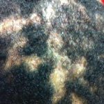
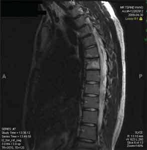
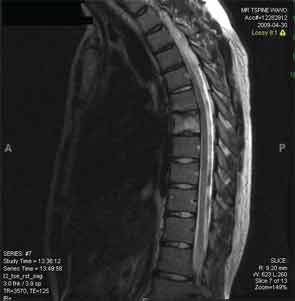
A 17-year-old male presented with pain in the thoracic spine and right medial clavicle and skin lesions on his palms and soles, previously diagnosed as pustular psoriasis. A biopsy of the sternum, performed by another physician who was concerned about infection, demonstrated a chronic osteomyelitis without granuloma formation. All microbial culture studies were negative. Magnetic resonance imaging (MRI) of the thoracic spine showed increased uptake at the T7 vertebra (see Figure 1). A biopsy of this bone showed marrow fibrosis with plasma cells present and rare lymphocytes. The microbial cultures were sterile. Treatment with oral and subsequently parenteral bisphosphonates and etanercept were all ineffective. He was then treated with infliximab for several months with considerable symptomatic relief and resolution of the MRI abnormality previously seen at T7 (see Figure 2).

Over time, despite progressive increases in the dose of infliximab, he continued to complain of significant pain. He was then treated with golimumab 50 mg monthly with some improvement. Phototherapy was prescribed for his skin lesions. Although the skin lesions improved, after eight months, the back pain worsened. The patient was switched to adalimumab 40 mg every other week. He continues to require narcotic analgesics for pain relief.

Patient 2

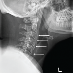
A 59-year-old woman developed severe pain in the thoracic spine and shoulder. A radiograph of the thoracic spine demonstrated a suspicious lesion at the T7 level. Biopsy of the lesion demonstrated nonspecific findings, and microbial cultures were negative. A biopsy of the rash over the palms was consistent with a neutrophilic pustulosis. Treatment was initiated with ciprofloxacin and trimethoprim/sulfamethoxazole. However, the patient developed disabling anterior chest pain for which treatment with several doses of intravenous pamidronate was temporarily helpful.

Page: 1 2 3 4 5 | Single Page
Share: 

Filed under:Clinical Criteria/GuidelinesConditionsOsteoarthritis and Bone Disorders Tagged with:anti-inflammatorybonecase reportimaginginfliximabPainpatient careradiographrheumatologistSAPHOsynovitisTreatment

Related Articles

    Case Report: A Patient Presents with Rare, Fulminant SAPHO Syndrome

    October 18, 2018

    Synovitis, acne, pustulosis, hyperostosis and osteitis (SAPHO) syndrome is a heterogeneous, inflammatory, musculoskeletal disease. The disease is an insidious, sterile osteitis with associated skin and synovial inflammation.1 Diagnosis can prove challenging, but a thorough clinical history, high clinical suspicion and imaging techniques can help clinch it. The below case reveals a rare, fulminant presentation of…

    Case Report: Acne Drug Leads Patient to Retinoid Hyperostosis

    November 19, 2018

    Presentation A 26-year-old man with a history of acne vulgaris and hidradenitis suppurativa presented to our rheumatology clinic with persistent back pain and stiffness of three years’ duration. He described bilateral low back pain that was worse when he arose in the morning and at night when he was trying to sleep. In a similar…

    Diagnostic Drill-Down: New Research Suggests Genetic Autoinflammatory Roots

    January 25, 2021

    Two experts discussed ongoing difficulties in diagnosing autoinflammatory disease, & promising new studies that point to possible genetic roots of autoinflammatory disease.

    Imaging in Ankylosing Spondylitis

    April 1, 2015

    MRI inflammation, fat and new bone formation in the sacroiliac joints, spine in patients with AS

  • About Us
  • Meet the Editors
  • Issue Archives
  • Contribute
  • Advertise
  • Contact Us
  • Copyright © 2025 by John Wiley & Sons, Inc. All rights reserved, including rights for text and data mining and training of artificial technologies or similar technologies. ISSN 1931-3268 (print). ISSN 1931-3209 (online).
  • DEI Statement
  • Privacy Policy
  • Terms of Use
  • Cookie Preferences