NEW YORK (Reuters Health)—Dual energy computed tomography (DECT) can differentiate cardiovascular monosodium urate (MSU) deposits from calcium deposits in patients with gout, potentially identifying those at risk of heart disease, researchers say. Sylvia Strobl, MD, of Medical University Innsbruck and colleagues analyzed calcium scores and MSU deposits in 59 patients with gout (mean age: 59;…

Catch Your Breath: Insights into ILD in RA Patients
Detecting interstitial lung disease in RA patients can be challenging. But evaluating risk factors and the use of imaging can help clinicians identify and manage this condition in patients…

Rheumatoid Arthritis Research Advances
SAN DIEGO—In a roundup of current research in rheumatoid arthritis (RA) presented at the 2017 ACR/ARHP Annual Meeting Nov. 3–8, Mark C. Genovese, MD, professor of medicine in the Division of Immunology and Rheumatology at Stanford University, Palo Alto, Calif., urged his audience to reflect on the impact therapy advances have made on RA. “In…
Predictive Value of Imaging Studied for Calcium Crystal Deposition in Rheumatic Diseases
MADRID—Calcification in osteoarthritis (OA) involves a series of pathways and interactions that feed off each other in a process that bears some resemblance to the transformation of cartilage to bone that takes place in the embryonic stage of human development, a researcher said here at the 2017 Annual European Congress on Rheumatology (EULAR). “My hypothesis…
Calcium Crystal Deposition in Rheumatic Diseases: Mechanisms & Evaluation of Calcium Crystal Deposits Explored
MADRID—Calcification in osteoarthritis (OA) involves a series of pathways and interactions that feed off each other in a process that bears some resemblance to the transformation of cartilage to bone that takes place in the embryonic stage of human development, a researcher said here at the 2017 Annual European Congress on Rheumatology (EULAR). “My hypothesis…
Predictive Value of Whole-Body MRI & Ultrasound Explored in EULAR Studies
Highlights from the 2017 EULAR Congress MADRID—Researchers say that whole-body MRI could yield an earlier diagnosis of spondyloarthropathy (SpA) in patients with early inflammatory joint symptoms, according to findings presented in a poster session at the Annual European Congress of Rheumatology (EULAR). The approach could lead to earlier treatment and better outcomes, they say. Investigators…
Ultrasound Can be Useful in Diagnosing Gout
The presence of synovial monosodium urate monohydrate (MSU) crystals is the gold standard for diagnosing gout. But a new study, funded in part by the ACR and led by rheumatologists, including Alexis Ogdie, MD, MSCE, evaluated the effectiveness of ultrasound in diagnosing it. The study found that ultrasound can be useful in discriminating gout from non-gout….
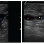
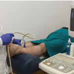
Rheumatology Case Report: Deep Vein Thrombosis Detected by Point-of-Care Ultrasound
Case A 46-year-old Caucasian female presented to the outpatient rheumatology clinic where she had been followed for several years. Her chief complaint was pain in her right knee, posterior right thigh and right hip that had begun gradually over the previous three weeks. Her past medical history was significant for rheumatoid arthritis (RA), obesity and…
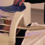
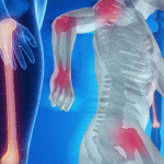
fMRI Can Help Diagnose Fibromyalgia
Brain imaging can distinguish fibromyalgia patients from healthy controls with high sensitivity and specificity, according to two papers published nearly simultaneously in Pain late last summer, by groups at the Universities of Colorado and Michigan, respectively. Somewhat surprisingly to the authors and others, in the Colorado study, which used both painful and nonpainful stimuli, the…
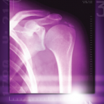
Ultrasound May Be Useful for Grading Rotator Cuff Tendinopathy
Researchers have developed procedures and assessed their efficacy for the use of ultrasound images to measure the inter-rater reliability of the measurement of structural changes in the tendon of patients with supraspinatus tendinopathy. The standardized procedures proved useful in evaluating patients…
- « Previous Page
- 1
- 2
- 3
- 4
- 5
- …
- 12
- Next Page »