A 66-year-old male patient presents to the office with right knee pain. He was in the office two weeks prior for a follow-up visit of his primary osteoarthritis. He received an injection of hyaluronate sodium in his right knee four months before and states that his knee felt like new. He states that everything was…

New Research into Rheumatoid Arthritis, Gout Includes Updates on Methotrexate, Biologics, Ultrasound
LONDON—From optimizing responses to methotrexate, to the efficacy of biologics, to the need for imaging in assessing remission, the literature, as ever, has been lively with explorations of pressing topics in the treatment and management of rheumatoid arthritis. Josef Smolen, MD, chair of rheumatology at the Medical University of Vienna, reviewed many of the highlights…
Ultrasound May Be Useful for Grading Rotator Cuff Tendinopathy
Researchers have developed procedures and assessed their efficacy for the use of ultrasound images to measure the inter-rater reliability of the measurement of structural changes in the tendon of patients with supraspinatus tendinopathy. The standardized procedures proved useful in evaluating patients…
From the Expert: Musculoskeletal Ultrasound Training Benefits Rheumatology Practices & Patients
Eugene Kissin, MD, says the use of musculoskeletal ultrasound can help rheumatologists diagnose and treat disease. Getting the proper training is critical. USSONAR is a training resource that can help…
Diagnostic Imaging in Lupus Patient with Foot Pain: Findings
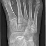
View the question. Findings/Diagnosis An anteroposterior (AP) radiograph of the right foot shows hallux valgus of the first metatarsal phalangeal (MTP) joint, erosive changes at the first and fifth metatarsal bones and degenerative changes at the fourth and fifth metatarsal-cuboid joints. An AP radiograph of the left foot shows extensive erosive and degenerative changes at…

Diagnostic Imaging in Lupus Patient with Foot Pain: History
Editor’s note: In this recurring feature, we first present a series of images (this page) for your review, and then a brief discussion of the findings and diagnosis. Before you turn to the discussion, examine these images carefully and draw your own conclusions. History A 33-year-old woman with a 16-year history of systemic lupus erythematosus…
Pulse-Echo Ultrasound Useful for Osteoporosis Screening
NEW YORK (Reuters Health)—Pulse-echo ultrasound is a useful method for point-of-care osteoporosis screening, researchers from Finland report. “To effectively increase diagnostic coverage, this kind of device should be in every primary or occupational healthcare unit,” Dr. Janne P. Karjalainen from the University of Eastern Finland in Kuopio tells Reuters Health by email. Currently, osteoporosis is…

Musculoskeletal Ultrasound: A Valuable Tool for Diagnosing Rheumatic Illnesses
Musculoskeletal (MSK) ultrasound is a valuable imaging modality for the practicing rheumatologist and provides an efficient tool with high diagnostic value in the evaluation of patients with musculoskeletal complaints. The use of MSK ultrasound has evolved in the U.S. due to the emergence of less-expensive, portable ultrasound units, which provide high-quality gray-scale and power Doppler…
EULAR 2015: Imaging in Rheumatology
ROME, Italy—The explosion of imaging technology has made it more important than ever to establish a standardized way in which imaging can and should be used in clinical practice, an expert said in a session at EULAR 2015, the annual congress of the European League Against Rheumatism (EULAR). Marie-Antonietta d’Agostino, MD, PhD, professor of rheumatology…
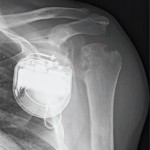
Diagnostic Imaging in Patient with Atraumatic Left Shoulder Pain: History
Editor’s note: In this recurring feature, we first present a series of images (this page) for your review, and then a brief discussion of the findings and diagnosis. Before you turn to the discussion, examine these images carefully and draw your own conclusions. History A 56-year-old man with end-stage renal disease on peritoneal dialysis presents…
- « Previous Page
- 1
- …
- 3
- 4
- 5
- 6
- 7
- 8
- Next Page »