It’s an exciting time for ultrasound in rheumatology, & it’s never too late to learn. Whether you’re just starting fellowship or have been practicing for decades, there’s a place for ultrasound in your practice.
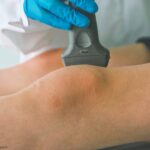
It’s an exciting time for ultrasound in rheumatology, & it’s never too late to learn. Whether you’re just starting fellowship or have been practicing for decades, there’s a place for ultrasound in your practice.
In a session on a current ACR project on musculoskeletal ultrasound (MSUS), Gurjit Kaeley, MBBS, RhMSUS, MRCP, division chief, professor of medicine, fellowship program director and program director for musculoskeletal ultrasound at the University of Florida College of Medicine, Jacksonville, will present information on proposed guidance related to rheumatoid arthritis. The ACR places a high…

The ACR places a high priority on developing relevant, practical guidance for the rheumatology community. A current ACR project on musculoskeletal ultrasound (MSUS) will be discussed in the ACR Convergence 2024 session, Proposed ACR Guidance for Use of Musculoskeletal Ultrasound in Rheumatoid and Psoriatic Arthritis, on Saturday, Nov. 16. In the session, Veena K. Ranganath,…
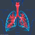
Shrinking lung syndrome (SLS) is a rare cause of dyspnea that has been most commonly described in patients with systemic lupus erythematosus (SLE), but is also found in systemic sclerosis, Sjögren’s disease and rheumatoid arthritis. Shrinking lung syndrome is characterized by a restrictive pattern on lung spirometry, despite normal lung parenchyma, and an elevated diaphragm.1…

A 17-year-old woman presents with chronic finger pain experienced over six months that is worse in the mornings. On physical exam, the patient has no joint swelling, pain on range of motion or limitation of range of motion in any of her finger joints. She has a tender, subcutaneous, firm, flesh-colored nodule on the lateral…
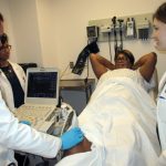
At the 17th Annual Advances in the Diagnosis & Treatment of the Rheumatic Diseases meeting, Dana DiRenzo, MD, MHS, RhMSUS, discussed the use of ultrasound imaging in patients with inflammatory arthritis.
Ultrasound may provide unique insights into the effects of immune checkpoint inhibitors on the human body beyond the immune system. Research suggests synovitis and inflammatory tendon involvement are commonly seen in patients with immune checkpoint inhibitor-induced inflammatory arthritis.
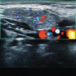
For most rheumatologists, the key elements of the physical exam—inspection, palpation, percussion and auscultation—have long been second nature, but a fifth modality has grown in importance with respect to making the correct diagnosis: ultrasound. From evaluating for Doppler signal and additional findings indicative of synovitis to identifying bony erosions, chondrocalcinosis, tophi and other articular and…
A 65-year-old woman was referred by an orthopedist to a rheumatologist for left knee pain. Previously, in 2014, she underwent left total knee arthroplasty (TKA) for severe osteoarthritis in a different institution. Following the procedure, she experienced severe chronic anterolateral knee pain at rest, exacerbated by walking. Because she was rendered wheelchair bound and required…

ATLANTA—Point-of-care ultrasound education mainly has occurred at the undergraduate level at U.S. medical schools, but rheumatology fellowship training programs are rapidly catching up and integrating it into their curricula, according to two program directors who reviewed the state of rheumatology ultrasound education, including potential barriers to its implementation, on Nov. 12 at the 2019 ACR/ARP…