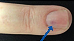
Figure 2: The nail bed shows some discoloration.
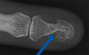
Figure 3: Erosion is seen at the distal phalanx.
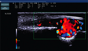
Figure 4: Color Doppler at the erosion site displays a 5 mm vascular lesion.
Case 3
A 33-year-old office manager presented with a two-year history of pain in the left hand and was referred for evaluation for possible median nerve entrapment at the wrist.
Electrodiagnostic studies and ultrasound of the carpal tunnel were normal. Her symptoms were most intense at the fourth fingernail bed, where she displayed acute tenderness to palpation and to pressure from the ultrasound probe. Discoloration under the nail (see Figure 5,) was apparent.
POCUS was performed at the symptomatic site, using a high-resolution 22 MHz ultrasound transducer. For the ultrasound assessment of superficial structures, such as the nail bed, a very high-frequency probe is required. (Note: In cases 2 and 3, a 22 MHz linear probe was used [see Figure 6]). A vascular malformation consistent with a 2 mm glomus tumor was suspected (see Figure 7). The ultrasound study guided the follow-up MRI examination, confirming an isolated glomus tumor (see Figure 8). The lesion was surgically excised.
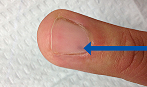
Figure 5: A splinter-shaped bluish discoloration under the nail bed.
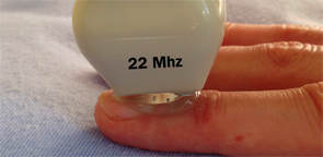
Figure 6: A high-frequency linear ultrasound probe (22 MHz) allows for very high-resolution imaging of superficial structures.
1 Year Later
All three patients report they are pain free one year after excisional therapy performed by a hand surgeon.
About Glomus Tumors
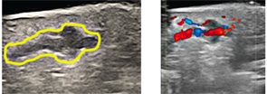
Figure 7: An irregular heterogeneous mass under nail on B Mode and a highly vascular discrete lesion on color Doppler.
Glomus tumors normally occur in tissues rich in glomus bodies: temperature- and pressure-sensitive receptors present in the stratum reticularis of the dermis. They represent 1–4% of all soft-tissue hand tumors. Roughly 70% of single tumors occur in patients younger than 30, and multiple tumors most often occur in patients between ages 15 and 20.2,3 Tumors can appear anywhere that glomoid cell types occur, and subungual glomus tumors are more common among women.2,4,5
As hamartomas, glomus tumors are formed from a hyperplasia of normal tissue.2,6 There are abundant polyhedral glomus tumor cells, seen as rounded uniform epithelioid cells with granular cytoplasm (see Figure 1). Staining with actin highlights smooth muscle surrounding vascular channels and neural elements (Figure 9).