The Rheumatologist
COVID-19 NewsACR Convergence
  • Connect with us:
  • Facebook
  • Twitter
  • LinkedIn
  • YouTube
  • Feed
  • Home
  • Conditions
    • Rheumatoid Arthritis
    • SLE (Lupus)
    • Crystal Arthritis
      • Gout Resource Center
    • Spondyloarthritis
    • Osteoarthritis
    • Soft Tissue Pain
    • Scleroderma
    • Vasculitis
    • Systemic Inflammatory Syndromes
    • Guidelines
  • Resource Centers
    • Axial Spondyloarthritis Resource Center
    • Gout Resource Center
    • Psoriatic Arthritis Resource Center
    • Rheumatoid Arthritis Resource Center
    • Systemic Lupus Erythematosus Resource Center
  • Drug Updates
    • Biologics & Biosimilars
    • DMARDs & Immunosuppressives
    • Topical Drugs
    • Analgesics
    • Safety
    • Pharma Co. News
  • Professional Topics
    • Ethics
    • Legal
    • Legislation & Advocacy
    • Career Development
      • Certification
      • Education & Training
    • Awards
    • Profiles
    • President’s Perspective
    • Rheuminations
    • Interprofessional Perspective
  • Practice Management
    • Billing/Coding
    • Quality Assurance/Improvement
    • Workforce
    • Facility
    • Patient Perspective
    • Electronic Health Records
    • Apps
    • Information Technology
    • From the College
    • Multimedia
      • Audio
      • Video
  • Resources
    • Issue Archives
    • ACR Convergence
      • Gout Resource Center
      • Axial Spondyloarthritis Resource Center
      • Psoriatic Arthritis
      • Abstracts
      • Meeting Reports
      • ACR Convergence Home
    • American College of Rheumatology
    • ACR ExamRheum
    • Research Reviews
    • ACR Journals
      • Arthritis & Rheumatology
      • Arthritis Care & Research
      • ACR Open Rheumatology
    • Rheumatology Image Library
    • Treatment Guidelines
    • Rheumatology Research Foundation
    • Events
  • About Us
    • Mission/Vision
    • Meet the Authors
    • Meet the Editors
    • Contribute to The Rheumatologist
    • Subscription
    • Contact
  • Advertise
  • Search
You are here: Home / Articles / A Duet of Bone and the Immune System

A Duet of Bone and the Immune System

July 12, 2011 • By Christian Beyer, MD, and Georg Schett, MD

  • Tweet
  • Email
Print-Friendly Version / Save PDF

In addition to mesenchymal cells, inflammatory cells—T lymphocytes in particular—are key elements that regulate osteoclastogenesis. Clinical observations suggest that osteoclastic bone resorption is associated with T lymphocyte activation in autoimmune and inflammatory disease. The role of T cells in osteoclastogenesis and bone metabolism is further supported by findings indicating that activated T lymphocytes express high levels of RANKL. The actual effect of T lymphocytes on bone, however, is modulated by their cytokine expression repertoire and their pattern of surface molecule expression.

You Might Also Like
  • Insight into Crosstalk Between Bone & Immune Systems
  • Th17 May Contribute to Bone Destruction by Modifying Osteoclasts
  • Methotrexate May Mitigate Inflammatory Bone Disease Note Experts at the ACR/ARHP Winter Rheumatology Symposium
Explore This Issue
July 2011

Although T cells can stimulate osteoclastogenesis, these cells also can display antiosteoclastogenic effects despite the expression of RANKL. This effect occurs particularly with TH1 cells expressing interferon-γ, a potent antiosteoclastogenic cytokine, and with regulatory T cells, which express high levels of CTLA4, a surface molecule important in inhibition of co-stimulation. In contrast, TH17 cells releasing IL-17 as well as TNF have been identified as important inducers of osteoclast differentiation.12 In this context, IL-17 may act in two ways. First, IL-17 can induce RANKL expression on mesenchymal cells; second, this cytokine causes local inflammation by the release of TNF, IL-17, and IL-22, further supporting RANKL expression.2,4

ad goes here:advert-1
ADVERTISEMENT
SCROLL TO CONTINUE

Immune-osteoblast Interactions

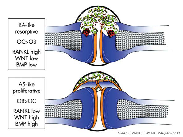
Although osteoclasts have been a major focus of attention in the study of inflammatory bone loss, surprisingly little is known about the activity of osteoblasts in this setting. Nevertheless, because bone metabolism depends on the balance between bone-resorbing osteoclasts and bone-forming osteoblasts, reduced bone formation likely contributes to bone loss in patients with arthritis. The mechanism behind this interaction is not fully defined, although TNF and other factors may repress bone formation, at least in part through the induction of dickkopf (Dkk)-1 and sclerostin, both inhibitors of the Wnt signaling, which is a central pathway for bone formation.13 Whereas Dkk-1 and sclerostin may act to suppress bone responses in diseases like rheumatoid arthritis (RA), impaired expression of these molecules also may be linked to aberrant bone formation, as found in spondyloarthritis (SpA). In addition to the effects of Wnt signaling, bone morphogenic proteins and transforming growth factor-β may induce new bone formation in arthritis.14 (See Figure 2, p. 37.)

Bone Erosion in RA

Figure 2: Joint remodelling in arthritis.
click for large version
Figure 2: Joint remodelling in arthritis. “RA-like” joint remodelling is based on the resorption of juxtaarticular bone by osteoclasts (OC, red cells). Molecules involved in osteoclast formation such as the receptor antagonist of NK-kB ligand (RANKL) dominate, whereas those important for osteoblasts (OB) and bone formation are suppressed. “AS-like” joint remodelling is based on bone formation at the joint ends leading to osteophytes, which are bony spurs, built by osteoblasts (orange cells). Molecules involved in osteoblast formation such as WNT and bone morphogenic proteins (BMP) dominate, whereas those important for osteoclasts and bone resorption are suppressed.

Inflammation is the primary trigger for the process of local bone erosions in RA, consistent with the prominence of inflammatory cell infiltrates in the tissue of patients with this disease (see Figure 3, at right). Bromley and Woolley and Gravallese and colleagues provided the first detailed descriptions of the location and activity of osteoclasts in the RA joint.15,16 These investigators demonstrated the expression of osteoclasts at the interface between advancing pannus tissue and the bone surface. Later, several groups demonstrated the essential role of osteoclasts in triggering inflammatory bone erosions in experimental disease models by blocking essential molecules for osteoclastogenesis or studying osteoclast-deficient mice.2,4

ad goes here:advert-2
ADVERTISEMENT
SCROLL TO CONTINUE

Pages: 1 2 3 4 5 6 | Single Page

Filed Under: Conditions, Systemic Inflammatory Syndromes Tagged With: Autoimmune disease, bone, Diagnostic Criteria, Immune System, Osteoimmunology, PathogenesisIssue: July 2011

You Might Also Like:
  • Insight into Crosstalk Between Bone & Immune Systems
  • Th17 May Contribute to Bone Destruction by Modifying Osteoclasts
  • Methotrexate May Mitigate Inflammatory Bone Disease Note Experts at the ACR/ARHP Winter Rheumatology Symposium
  • Clues Emerge to Bone Health, Destruction in Rheumatoid Arthritis

Patient & Caregiver Resources

Find a rheumatology provider. Learn about your condition and how to live with it. English and Spanish language resources.

View Patient & Caregiver Resources »

Rheumatology Research Foundation

The Foundation is the largest private funding source for rheumatology research and training in the U.S.

Learn more »

American College of Rheumatology

Visit the official website for the American College of Rheumatology.

Visit the ACR »

The Rheumatologist newsmagazine reports on issues and trends in the management and treatment of rheumatic diseases. The Rheumatologist reaches 11,500 rheumatologists, internists, orthopedic surgeons, nurse practitioners, physician assistants, nurses, and other healthcare professionals who practice, research, or teach in the field of rheumatology.

About Us / Contact Us / Advertise / Privacy Policy / Terms of Use / Cookie Preferences

  • Connect with us:
  • Facebook
  • Twitter
  • LinkedIn
  • YouTube
  • Feed

Copyright © 2006–2023 American College of Rheumatology. All rights reserved.

ISSN 1931-3268 (print)
ISSN 1931-3209 (online)