Shortly afterward, he was found to have moderately elevated transaminases and a greatly elevated alkaline phosphatase at 308 U/L (upper limit of normal 126 U/L). An abdominal ultrasound showed cirrhosis and interval development of multiple hypoechoic lesions throughout the liver, concerning for metastatic disease or hepatocellular carcinoma. Computerized tomography (CT) of the abdomen also demonstrated numerous ill-defined hypodense lesions all throughout the liver (see Figure 1), suspicious for metastatic disease, along with a 1.9×2.9 cm mesorectal mass concerning for colorectal cancer.
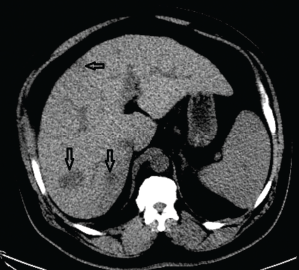
Figure 1. A CT of the abdomen demonstrated numerous hypodense lesions present in both lobes of the liver, with the largest lesion measuring 2.0 x 3.1 cm.
This was followed up by a magnetic resonance imaging (MRI) scan of his abdomen, which corroborated the previous hepatic findings. All lesions on the MRI were deemed stable since the earlier CT, and no new lesions were noted. We felt it would be unlikely for the hepatic lesions to be metastatic disease due to stable preexisting lesions and the lack of new lesions in this time frame. An ultrasound-guided biopsy was performed, which revealed necrotizing granulomatous disease (see Figure 2).
Given this granulomatous disease and history of polyarthralgia, sarcoidosis was considered. Plain films were updated, and an MRI of his hands was obtained to determine if his arthropathy and liver disease could be due to sarcoidosis; however, the MRI was remarkable only for lesions consistent with a crystalline arthropathy (see Figure 3). Serum angiotensin I-converting enzyme and vitamin D levels were also not supportive of sarcoidosis.
Infectious etiologies were high on the differential for his granulomatous disease, especially after it was noted our patient had a latent tuberculosis (TB) infection when he was a child in the 1980s. He had received at least one month of treatment for latent TB, but was unsure of any further details of his treatment. Acid-fast bacilli and Gomori methenamine silver stains of the biopsy specimen were negative.
An infectious disease specialist was subsequently consulted. The patient denied fevers or chills, but he reported fatigue and recent weight gain.
He had worked as a long-haul truck driver and had a military history with deployment to the Persian Gulf. Serologies obtained for cryptococcus, histoplasma and coccidioides were unremarkable. QuantiFERON-TB Gold was positive, not unexpected given his latent TB infection as a child. A repeat liver biopsy found granulomatous inflammation with focal necrosis in a background of features compatible with cirrhosis, consistent with previous biopsy. The acid-fast bacilli smear was again negative.