Video: Every Case Tells a Story| Webinar: ACR/CHEST ILD Guidelines in Practice

An official publication of the ACR and the ARP serving rheumatologists and rheumatology professionals

  • Conditions
    • Axial Spondyloarthritis
    • Gout and Crystalline Arthritis
    • Myositis
    • Osteoarthritis and Bone Disorders
    • Pain Syndromes
    • Pediatric Conditions
    • Psoriatic Arthritis
    • Rheumatoid Arthritis
    • Sjögren’s Disease
    • Systemic Lupus Erythematosus
    • Systemic Sclerosis
    • Vasculitis
    • Other Rheumatic Conditions
  • FocusRheum
    • ANCA-Associated Vasculitis
    • Axial Spondyloarthritis
    • Gout
    • Psoriatic Arthritis
    • Rheumatoid Arthritis
    • Systemic Lupus Erythematosus
  • Guidance
    • Clinical Criteria/Guidelines
    • Ethics
    • Legal Updates
    • Legislation & Advocacy
    • Meeting Reports
      • ACR Convergence
      • Other ACR meetings
      • EULAR/Other
    • Research Rheum
  • Drug Updates
    • Analgesics
    • Biologics/DMARDs
  • Practice Support
    • Billing/Coding
    • EMRs
    • Facility
    • Insurance
    • QA/QI
    • Technology
    • Workforce
  • Opinion
    • Patient Perspective
    • Profiles
    • Rheuminations
      • Video
    • Speak Out Rheum
  • Career
    • ACR ExamRheum
    • Awards
    • Career Development
  • ACR
    • ACR Home
    • ACR Convergence
    • ACR Guidelines
    • Journals
      • ACR Open Rheumatology
      • Arthritis & Rheumatology
      • Arthritis Care & Research
    • From the College
    • Events/CME
    • President’s Perspective
  • Search

Case Report: A Rare Mimic of Giant Cell Arteritis

Iryna Nemesh, MD, Saleema Kherani, MD MPH, Shikha Singla, MD, & William Wirostko, MD  |  Issue: November 2021  |  November 14, 2021

Optos Fundus photo of the right eye with subtle area of retinal whitening in the macula with mild vitreous opacities inferiorly (arrowhead).

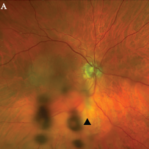
Figure 1A. Optos Fundus photo of the right eye with subtle area of retinal whitening in the macula with mild vitreous opacities inferiorly (arrowhead).

Optos Fundus photo of the left eye with large placoid area of retinal whitening involving the macula (arrow), with retinal hemorrhages and vitreous opacity temporally.

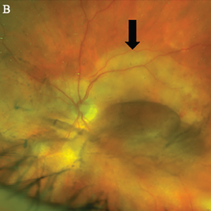
Figure 1B, Optos Fundus photo of the left eye with large placoid area of retinal whitening involving the macula (arrow), with retinal hemorrhages and vitreous opacity temporally.


He denied any eye pain, pain on eye movements, complete vision loss, new neurologic symptoms, fevers, chills, fatigue, weight loss or jaw claudication.

In the clinic, the initial fundoscopic exam appeared normal. Inflammatory markers were significantly elevated with a C-reactive protein (CRP) level of 35.8 mg/L and an erythrocyte sedimentation rate (ESR) of 130 mm/hour.

ad goes here:advert-1
ADVERTISEMENT
SCROLL TO CONTINUE

He was referred to the emergency department with a concern for giant cell arteritis.

On initial examination, patient was afebrile with stable hemodynamics. Bilateral temporal artery pulses were present and equal. There was no temporal artery nodularity or tenderness to palpation on either side.

ad goes here:advert-2
ADVERTISEMENT
SCROLL TO CONTINUE

Magnetic resonance imaging (MRI) of the brain showed chronic microvascular ischemic changes, but no changes in the orbit or optic nerve. He received pulse-dose steroids with 1,000 mg of intravenous (IV) methylprednisolone daily for three days for presumed GCA, and was continued on 60 mg of oral prednisone daily. A bilateral temporal artery biopsy was performed, but was unrevealing.

The patient said his right eye vision and right temple pain improved with steroids. Given the high clinical suspicion for GCA and the improvement in his symptoms, the decision was made to continue treatment with steroids.

The patient presented to the rheuma­tology clinic in follow-up a few weeks later. At the time of the visit, he reported a new-onset sharp pain and blurry vision in the left eye. He denied headaches at that time and had an unrevealing physical exam. Due to his new onset of visual symptoms despite being on high-dose steroids, as well as previously negative temporal artery biopsy results, GCA was presumed to be unlikely etiology and a steroid taper was initiated by the rheumatologist.

The patient was reevaluated by an ophthalmologist to address the new vision changes. Subsequent ophthalmologic exam showed bilateral placoid lesions involving the macula, characteristic for syphilitic placoid chorioretinitis (see Figures 1A&B and 2).

As a part of his broader workup, a test for Treponema pallidum was ordered. A plasma reagin (RPR) test was positive, with titers at 1:128. A fluorescent treponemal antibody absorption (FTA-Abs) test was also positive.

The patient was referred to an infectious disease specialist for further management. At the time of infectious disease visit, his sexual history was confirmed, and he reported a homosexual encounter with a new partner about three weeks prior to presentation. No protection was used at the time.

Further tests for infectious diseases were negative for HIV and hepatitis C. Based on his clinical presentation and high-risk sexual exposure, the diagnosis of ocular syphilis was made.

The patient had no other neurological complaints, so a decision to obtain lumbar puncture for cerebrospinal fluid (CSF) analysis was deferred. The patient completed a 14-day course of IV peni­cillin. He has been closely followed up by infectious disease and ophthalmology teams. His visual symptoms improved subjectively and objectively on follow-up with ophthalmology.

Page: 1 2 3 4 | Single Page
Share: 

Filed under:ConditionsVasculitis Tagged with:ocular diseasesyphilis

Related Articles

    Two Inflammatory Conditions—Polymyalgia Rheumatica and Giant Cell Arteritis—Share Clinical Connection

    March 1, 2013

    Polymyalgia rheumatica (PMR) and giant cell arteritis (GCA) have common clinical and epidemiologic links, but they need not occur synchronously

    Atypical Giant Cell Arteritis Case Illustrates Diagnosis, Management Challenges

    Atypical Giant Cell Arteritis Case Illustrates Diagnosis, Management Challenges

    November 14, 2021

    Giant cell arteritis (GCA) is a granulomatous vasculitis of large- and medium-sized arteries, usually affecting the cranial branches of the aortic arch. It is the most common vasculitis, with the highest risk factor being age. Accurate diagnosis and prompt initiation of therapy are of great importance to prevent serious complications, with the most feared being…

    Experts Discuss Proposed Giant Cell Arteritis Risk Tool

    April 26, 2018

    A proposed model to predict the risk of giant cell arteritis (GCA) prior to a temporal artery biopsy could help triage patients and guide decision making about the need for biopsy or monitoring (see Figure 1). There’s no specific biomarker for GCA, and GCA can be a “diagnostic conundrum, especially when it presents in an…

    Updates on Giant Cell Arteritis

    March 19, 2018

    SAN DIEGO—Recent research tells us more about giant cell arteritis (GCA) to help rheumatologists more accurately diagnose and effectively treat patients with this type of vasculitis. On Nov. 6 at the ACR/ARHP Annual Meeting, three experts explored the latest findings on GCA pathogenesis, diagnostic approaches, imaging modalities and growing treatment options. GCA: What’s Really Happening?…

  • About Us
  • Meet the Editors
  • Issue Archives
  • Contribute
  • Advertise
  • Contact Us
  • Copyright © 2025 by John Wiley & Sons, Inc. All rights reserved, including rights for text and data mining and training of artificial technologies or similar technologies. ISSN 1931-3268 (print). ISSN 1931-3209 (online).
  • DEI Statement
  • Privacy Policy
  • Terms of Use
  • Cookie Preferences