Video: Every Case Tells a Story| Webinar: ACR/CHEST ILD Guidelines in Practice

An official publication of the ACR and the ARP serving rheumatologists and rheumatology professionals

  • Conditions
    • Axial Spondyloarthritis
    • Gout and Crystalline Arthritis
    • Myositis
    • Osteoarthritis and Bone Disorders
    • Pain Syndromes
    • Pediatric Conditions
    • Psoriatic Arthritis
    • Rheumatoid Arthritis
    • Sjögren’s Disease
    • Systemic Lupus Erythematosus
    • Systemic Sclerosis
    • Vasculitis
    • Other Rheumatic Conditions
  • FocusRheum
    • ANCA-Associated Vasculitis
    • Axial Spondyloarthritis
    • Gout
    • Psoriatic Arthritis
    • Rheumatoid Arthritis
    • Systemic Lupus Erythematosus
  • Guidance
    • Clinical Criteria/Guidelines
    • Ethics
    • Legal Updates
    • Legislation & Advocacy
    • Meeting Reports
      • ACR Convergence
      • Other ACR meetings
      • EULAR/Other
    • Research Rheum
  • Drug Updates
    • Analgesics
    • Biologics/DMARDs
  • Practice Support
    • Billing/Coding
    • EMRs
    • Facility
    • Insurance
    • QA/QI
    • Technology
    • Workforce
  • Opinion
    • Patient Perspective
    • Profiles
    • Rheuminations
      • Video
    • Speak Out Rheum
  • Career
    • ACR ExamRheum
    • Awards
    • Career Development
  • ACR
    • ACR Home
    • ACR Convergence
    • ACR Guidelines
    • Journals
      • ACR Open Rheumatology
      • Arthritis & Rheumatology
      • Arthritis Care & Research
    • From the College
    • Events/CME
    • President’s Perspective
  • Search

Case Report: Abscess as a Manifestation of Autoinflammatory Disease

Katherine Chakrabarti, MD, & Andrew Vreede, MD  |  Issue: June 2022  |  June 14, 2022

Axial CT showing multilocular abdominal fluid collection.

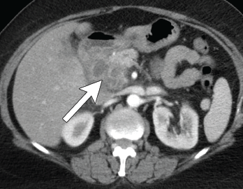
Figure 4. Axial CT showing multilocular abdominal fluid collection.

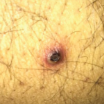
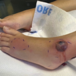
A 56-year-old woman presented to the hospital with chest and abdominal pain. Her history was notable for pustular psoriasis, psoriatic arthritis (untreated), relapsing-remitting multiple sclerosis (not on therapy), as well as recurrent abscesses over the preceding four years. Abscesses occurred at the chest wall (see Figures 2 & 3, opposite), axilla and pancreas.

She was treated with systemic antibiotics, although aerobic, anaerobic and AFB cul­tures were repeatedly negative during each of these episodes. Additional testing included negative results for anti-nuclear antibody, anti-neutrophil cytoplasmic antibody, anti-myeloperoxidase antibody, anti-proteinase 3 antibody and human leukocyte antigen B27. Computerized tomography (CT) of the abdomen/pelvis demonstrated a possible duodenal-colonic fistula that was not further evaluated. Her symptoms improved with corticosteroids.

ad goes here:advert-1
ADVERTISEMENT
SCROLL TO CONTINUE

One year later she was admitted with chest and abdominal pain. She was afebrile, hypertensive up to 200/110 mmHg (RR: <120 mmHg systolic, <80 mmHg diastolic), without tachycardia or abnormal respiratory rate. Initial labs were notable for leuko­cytosis with a total WBC of 22.2 x 10⁹/L (RR: 4.0–11.0 x 10⁹/L) and elevated inflammatory markers with ESR 82 mm/h (RR: 0–30 mm/hour for women over 50) and a CRP of 10.6 mg/dL (RR: 0–0.6 mg/dL). CT scan of the chest/abdomen/pelvis demonstrated a 2.3×4 cm peri-pancreatic abscess with fat stranding and associated lymphadenopathy (see Figures 4 & 5, p. 22).

Coronal CT showing multilocular abdominal fluid collection.

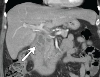
Figure 5. Coronal CT showing multilocular abdominal fluid collection.

Cultures from the abscess aspirate again failed to identify an organism, and 40 mg of prednisone daily was initiated. Despite this, she developed a new chest wall abscess over the following days. Incision and drainage were performed, with negative cultures. The following day she developed skin erythema, and pain and warmth at the incision and drainage sites. Due to concern for possible cellulitis, corticosteroids were stopped and she was started on antibiotics.

ad goes here:advert-2
ADVERTISEMENT
SCROLL TO CONTINUE

Ultimately, however, a new chest wall abscess developed, prompting re-initiation of 40 mg of prednisone daily. Her chest wall lesions improved over the following days. Due to suspicion of possible under­lying IBD, a fecal calprotectin was tested but proved indeterminate at 106 mg/kg (negative: <50 mg/kg). Colonoscopy was sub­sequently performed and was unremarkable.

A diagnosis of synovitis, acne, pustulosis, hyperostosis, osteitis syndrome (SAPHO) was considered given her chest wall lesions and psoriasis, but she lacked other clinical features of osteitis, hyperostosis or acne. She was diagnosed with spondyloarthropathy-associated aseptic abscess syndrome. She was discharged on a prednisone taper and subsequently lost to follow-up.

Discussion

Aseptic abscesses were first described in 1995 in the case of a 25-year-old man who developed multiple abscesses several years prior to an eventual diagnosis of Crohn’s disease.1 In 2007, the most complete description of this new entity was published with the inclusion of 30 cases from a French registry and 19 cases compiled from a literature review.2 The researchers defined AA by the radiographic appearance of an abscess, negative microbial diagnostic studies, lack of response to antibiotic therapy and rapid response to corticosteroids (see Table 1).2

Table 1: The 5 Features of Aseptic Abscesses

1. Presence of abscess on radiologic examination
2. Histopathology with neutrophilic features
3. Negative blood cultures and infectious evaluation
4. Failure of antibiotic therapy (defined as >2 weeks of therapy or >3 months if
concern for tuberculosis)
5. Improvement with steroids

Page: 1 2 3 4 | Single Page
Share: 

Filed under:Conditions Tagged with:abscessaseptic abscesscase reportSAPHO

Related Articles

    Case Report: Possible Overlapping Vasculitis & Ulcerative Colitis

    January 17, 2020

    A 42-year-old man with a history of ulcerative colitis (UC), primary sclerosing cholangitis (PSC) and chronic sinusitis was referred to a rheumatologist to evaluate for a possible diagnosis of systemic vasculitis. This patient had developed new skin lesions, gingival hypertrophy and ulcerating tracheobronchitis, concerning for possible granulomatosis with polyangiitis (GPA). Since 1994, the patient had…

    Case Report: Sweet Syndrome as an Initial Presentation of Crohn’s Disease

    July 13, 2022

    Acute febrile neutrophilic dermatosis, or Sweet syndrome, is an inflammatory disease that classically presents with fever, leukocytosis and tender, erythematous plaques characterized by neutrophilic infiltrates on biopsy. Sweet syndrome has been reported in association with several autoimmune diseases, including inflammatory bowel disease, systemic lupus erythematous, rheumatoid arthritis and sarcoidosis.1 Here, we discuss a case of…

    Case Report: A Patient Presents with Rare, Fulminant SAPHO Syndrome

    October 18, 2018

    Synovitis, acne, pustulosis, hyperostosis and osteitis (SAPHO) syndrome is a heterogeneous, inflammatory, musculoskeletal disease. The disease is an insidious, sterile osteitis with associated skin and synovial inflammation.1 Diagnosis can prove challenging, but a thorough clinical history, high clinical suspicion and imaging techniques can help clinch it. The below case reveals a rare, fulminant presentation of…

    What Rheumatologists Should Look for in Diagnosing SAPHO Syndrome

    November 1, 2012

    SAPHO syndrome (synovitis, acne, pustulosis, hyperostosis, and osteitis) is an obscure condition that can vex patients and clinicians

  • About Us
  • Meet the Editors
  • Issue Archives
  • Contribute
  • Advertise
  • Contact Us
  • Copyright © 2025 by John Wiley & Sons, Inc. All rights reserved, including rights for text and data mining and training of artificial technologies or similar technologies. ISSN 1931-3268 (print). ISSN 1931-3209 (online).
  • DEI Statement
  • Privacy Policy
  • Terms of Use
  • Cookie Preferences